Pleural effusion
Fluid in the chest; Fluid on the lung; Pleural fluid
A pleural effusion is a buildup of fluid between the layers of tissue that line the lungs and chest cavity.
Causes
The body produces pleural fluid in small amounts to lubricate the surfaces of the pleura. This is the thin tissue that lines the chest cavity and surrounds the lungs. Pleural effusion is an abnormal, excessive collection of this fluid.
There are two types of pleural effusion:
-
Transudative pleural effusion is caused by fluid leaking into the pleural space. This is from increased pressure in the blood vessels or a low blood protein count.
Heart failure
is the most common cause.
Heart failure
Heart failure is a condition in which the heart is no longer able to pump oxygen-rich blood to the rest of the body efficiently. This causes symptom...
 ImageRead Article Now Book Mark Article
 - Exudative effusion is caused by blocked blood vessels or lymph vessels, inflammation, lung injury, and tumors.
Risk factors of pleural effusion may include:
- Smoking and drinking alcohol
- Any previous complaint of high blood pressure
- History of any contact with asbestos
Symptoms
Symptoms can include any of the following:
-
Chest pain
, usually a sharp pain that is worse with cough or deep breaths
Chest pain
Chest pain is discomfort or pain that you feel anywhere along the front of your body between your neck and upper abdomen.
 ImageRead Article Now Book Mark Article
 -
Cough
Cough
Coughing is an important way to keep your throat and airways clear. But too much coughing may mean you have a disease or disorder. Some coughs are d...
 ImageRead Article Now Book Mark Article
 - Fever and chills
-
Hiccups
Hiccups
A hiccup is an unintentional movement (spasm) of the diaphragm, the muscle at the base of the lungs. The spasm is followed by quick closing of the v...
Read Article Now Book Mark Article -
Rapid breathing
Rapid breathing
Hyperventilation is rapid and deep breathing. It is also called overbreathing, and it may leave you feeling breathless.
Read Article Now Book Mark Article -
Shortness of breath
Shortness of breath
Breathing difficulty may involve:Difficult breathingUncomfortable breathingFeeling like you are not getting enough air
 ImageRead Article Now Book Mark Article
Sometimes there are no symptoms.
Exams and Tests
Your health care provider will examine you and ask about your symptoms. The provider will also listen to your lungs with a stethoscope and tap (percuss) your chest and upper back.
Chest CT scan or a chest x-ray may be enough for your provider to decide on treatment.
Chest CT scan
A chest CT (computed tomography) scan is an imaging method that uses x-rays to create cross-sectional pictures of the chest and upper abdomen....

Chest x-ray
A chest x-ray is an x-ray of the chest, lungs, heart, large arteries, ribs, and diaphragm.

Your provider may want to perform tests on the fluid. If so, a sample of fluid is removed with a needle inserted between the ribs. Tests on the fluid will be done to look for:
Fluid is removed with a needle
Thoracentesis is a procedure to remove fluid from the space between the lining of the outside of the lungs (pleura) and the wall of the chest....
- Infection
- Cancer cells
- Protein levels
Blood tests that may be done include
- Complete blood count (CBC), to check for signs of infection or anemia
- Kidney and liver function blood tests
If needed, these other tests may be done:
- Ultrasound of the heart (echocardiogram) to look for heart failure
- Lung biopsy to look for cancer
-
Passing a tube through the windpipe to check the airways for problems or cancer (
bronchoscopy
)
Bronchoscopy
Bronchoscopy is a test to view the airways and diagnose lung disease. It may also be used during the treatment of some lung conditions.
 ImageRead Article Now Book Mark Article
Treatment
The goal of treatment is to:
- Remove the fluid
- Prevent fluid from building up again
- Determine and treat the cause of the fluid buildup
Removing the fluid (thoracentesis) may be done if there is a lot of fluid and it is causing chest pressure, shortness of breath, or a low oxygen level. Removing the fluid allows the lung to expand, making breathing easier.
The cause of the fluid buildup must also be treated:
- If it is due to heart failure, you may receive diuretics (water pills) and other medicines to treat heart failure.
- If it is due to an infection, antibiotics will be given.
In people with cancer or infection, the effusion is often treated by using a chest tube to drain the fluid.
In some cases, any of the following treatments are done:
-
Chemotherapy
Chemotherapy
The term chemotherapy is used to describe cancer-killing drugs. Chemotherapy may be used to:Cure the cancerShrink the cancerPrevent the cancer from ...
 ImageRead Article Now Book Mark Article
 - Placing medicine into the chest that prevents fluid from building up again after it is drained
-
Radiation therapy
Radiation therapy
Radiation therapy uses high-powered x-rays, particles, or radioactive seeds to kill cancer cells.
 ImageRead Article Now Book Mark Article
 - Surgery
Outlook (Prognosis)
The outcome depends on the underlying disease.
Possible Complications
Complications of pleural effusion may include:
- Lung damage
-
Infection that turns into an abscess, called an
empyema
Empyema
Empyema is a collection of pus in the space between the lung and the inner surface of the chest wall (pleural space).
 ImageRead Article Now Book Mark Article
 -
Air in the chest cavity (
pneumothorax
) after drainage of the effusion
Pneumothorax
A collapsed lung occurs when air escapes from the lung. The air then fills the space outside of the lung, between the lung and chest wall. This bui...
 ImageRead Article Now Book Mark Article
 - Pleural thickening (scarring of the lining of the lung)
When to Contact a Medical Professional
Call your provider or go to the emergency room if you have:
- Symptoms of pleural effusion
- Shortness of breath or difficulty breathing right after thoracentesis
References
Broaddus VC, Light RW. Pleural effusion. In: Broaddus VC, Mason RJ, Ernst JD, et al, eds. Murray and Nadel's Textbook of Respiratory Medicine . 6th ed. Philadelphia, PA: Elsevier Saunders; 2016:chap 79.
Mccool FD. Diseases of the diaphragm, chest wall, pleura and mediastinum. In: Goldman L, Schafer AI, eds. Goldman-Cecil Medicine . 25th ed. Philadelphia, PA: Elsevier Saunders; 2016:chap 99.
-
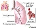
Lungs - illustration
The major features of the lungs include the bronchi, the bronchioles and the alveoli. The alveoli are the microscopic blood vessel-lined sacks in which oxygen and carbon dioxide gas are exchanged.
Lungs
illustration
-
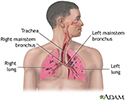
Respiratory system - illustration
Air is breathed in through the nasal passageways, travels through the trachea and bronchi to the lungs.
Respiratory system
illustration
-
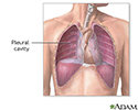
Pleural cavity - illustration
The pleural cavity is composed of the layers of the membrane lining the lung and the chest cavity.
Pleural cavity
illustration
-
Lungs - illustration
The major features of the lungs include the bronchi, the bronchioles and the alveoli. The alveoli are the microscopic blood vessel-lined sacks in which oxygen and carbon dioxide gas are exchanged.
Lungs
illustration
-
Respiratory system - illustration
Air is breathed in through the nasal passageways, travels through the trachea and bronchi to the lungs.
Respiratory system
illustration
-
Pleural cavity - illustration
The pleural cavity is composed of the layers of the membrane lining the lung and the chest cavity.
Pleural cavity
illustration
Review Date: 8/21/2016
Reviewed By: Denis Hadjiliadis, MD, MHS, Paul F. Harron, Jr. Associate Professor of Medicine, Pulmonary, Allergy, and Critical Care, Perelman School of Medicine, University of Pennsylvania, Philadelphia, PA. Also reviewed by David Zieve, MD, MHA, Isla Ogilvie, PhD, and the A.D.A.M. Editorial team.


