Ear wax
Ear impaction; Cerumen impaction; Ear blockage; Hearing loss - ear wax
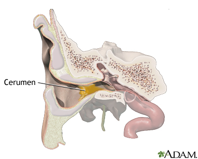
The ear canal is lined with hair follicles. The ear canal also has glands that produce a waxy oil called cerumen. The wax will most often make its way to the opening of the ear. There it will fall out or be removed by washing.
Wax can build up and block the ear canal. Wax blockage is one of the most common causes of hearing loss.
Causes
Ear wax protects the ear by:
- Trapping and preventing dust, bacteria, and other germs and small objects from entering and damaging the ear
- Protecting the delicate skin of the ear canal from getting irritated when water is in the canal
In some people, the glands produce more wax than can be easily removed from the ear. This extra wax may harden in the ear canal and block the ear. When you try to clean the ear, you may instead push wax deeper and block the ear canal.
Symptoms
Some of the common symptoms are:
-
Earache
Earache
An earache is a sharp, dull, or burning pain in one or both ears. The pain may last a short time or be ongoing. Related conditions include:Otitis m...
 ImageRead Article Now Book Mark Article
 - Fullness in the ear or a sensation that the ear is plugged
-
Noises in the ear (
tinnitus
)
Tinnitus
Tinnitus is the medical term for "hearing" noises in your ears. It occurs when there is no outside source of the sounds. Tinnitus is often called "r...
 ImageRead Article Now Book Mark Article
 - Partial hearing loss, may get worse
Treatment
Most cases of ear wax blockage can be treated at home. The following remedies can be used to soften wax in the ear:
- Baby oil
- Commercial drops
- Glycerin
- Mineral oil
- Water
Another method is to wash out the wax.
-
Use body-temperature water (cooler or warmer water may cause brief but severe dizziness or
vertigo
).
Vertigo
Dizziness is a term that is often used to describe 2 different symptoms: lightheadedness and vertigo. Lightheadedness is a feeling that you might fai...
 ImageRead Article Now Book Mark Article
 - Hold your head upright and straighten the ear canal by holding the outside ear and gently pulling upward.
- Use a syringe (you can buy one at the store) to gently direct a small stream of water against the ear canal wall next to the wax plug.
- Tip your head to allow the water to drain. You may need to repeat irrigation several times.
To avoid damaging your ear or causing an infection:
- Never irrigate the ear if the eardrum may have a hole in it.
- Do not irrigate the ear with a jet irrigator designed for cleaning teeth (such as a WaterPik).
After the wax is removed, dry the ear thoroughly. You may use a few drops of alcohol in the ear or a hair dryer set on low to help dry the ear.
You may clean the outer ear canal by using a cloth or paper tissue wrapped around your finger. Mineral oil can be used to moisturize the ear and prevent the wax from drying.
Do not clean your ears too often or too hard. Ear wax also helps protect your ears. Never try to clean the ear by putting any object, such as a cotton swab, into the ear canal.
If you cannot remove the wax plug or you have discomfort, consult a health care provider, who may remove the wax by:
- Repeating the irrigation attempts
- Suctioning the ear canal
- Using a small device called a curette
- Using a microscope to help
Outlook (Prognosis)
The ear may become blocked with wax again in the future. Hearing loss is often temporary. In most cases, hearing returns completely after the blockage is removed.
Rarely, trying to remove ear wax may cause an infection in the ear canal. This can also damage the eardrum.
When to Contact a Medical Professional
See your provider if your ears are blocked with wax and you are unable to remove the wax.
Also call if you have an ear wax blockage and you develop new symptoms, such as:
-
Drainage from the ear
Drainage from the ear
Ear discharge is drainage of blood, ear wax, pus, or fluid from the ear.
 ImageRead Article Now Book Mark Article
 -
Ear pain
Ear pain
An earache is a sharp, dull, or burning pain in one or both ears. The pain may last a short time or be ongoing. Related conditions include:Otitis m...
 ImageRead Article Now Book Mark Article
 -
Fever
Fever
Fever is the temporary increase in the body's temperature in response to a disease or illness. A child has a fever when the temperature is at or abov...
 ImageRead Article Now Book Mark Article
 -
Hearing loss
that continues after you clean the wax
Hearing loss
Hearing loss is being partly or totally unable to hear sound in one or both ears.
 ImageRead Article Now Book Mark Article
References
Lee DJ, Roberts D. Topical therapies for external ear disorders. In: Flint PW, Haughey BH, Lund V, et al, eds. Cummings Otolaryngology . 6th ed. Philadelphia, PA: Elsevier Saunders; 2015:chap 138.
O'Handley JG, Tobin EJ, Shah AR. Otorhinolaryngology. In: Rakel RE, Rakel DP, eds. Textbook of Family Medicine . 9th ed. Philadelphia, PA: Elsevier; 2016:chap 18.
Pfaff JA, Moore GP. Otolaryngology. In: Marx JA, Hockberger RS, Walls RM, eds. Rosen's Emergency Medicine . 8th ed. Philadelphia, PA: Elsevier Saunders; 2014:chap 72.
Riviello RJ. Otolaryngologic procedures. In: Roberts JR, ed. Roberts and Hedges' Clinical Procedures in Emergency Medicine . 6th ed. Philadelphia, PA: Elsevier Saunders; 2014:chap 63.
Roland PS, Smith TL, Schwartz SR, et al. Clinical practice guideline: cerumen impaction. Otolaryngol Head Neck Surg . 2008;139(3 Suppl 2):S1-S21. PMID: 18707628. www.ncbi.nlm.nih.gov/pubmed/18707628 .
-
Tips on removing ear wax
Animation
-
Wax blockage in the ear - illustration
The ear canal is lined with hair follicles and glands that produce a waxy oil called cerumen. Sometimes the glands produce more wax than can be easily excreted out the ear. This extra wax may harden within the ear canal and block the ear.
Wax blockage in the ear
illustration
-
Ear anatomy - illustration
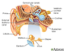
The ear consists of external, middle, and inner structures. The eardrum and the 3 tiny bones conduct sound from the eardrum to the cochlea.
Ear anatomy
illustration
-
Medical findings based on ear anatomy - illustration
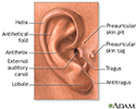
The external structures of the ear may aid in diagnosing some conditions by the presence or absence of normal landmarks and abnormal features including: earlobe creases, preauricular pits, and preauricular tags.
Medical findings based on ear anatomy
illustration
-
Wax blockage in the ear - illustration
The ear canal is lined with hair follicles and glands that produce a waxy oil called cerumen. Sometimes the glands produce more wax than can be easily excreted out the ear. This extra wax may harden within the ear canal and block the ear.
Wax blockage in the ear
illustration
-
Ear anatomy - illustration
The ear consists of external, middle, and inner structures. The eardrum and the 3 tiny bones conduct sound from the eardrum to the cochlea.
Ear anatomy
illustration
-
Medical findings based on ear anatomy - illustration
The external structures of the ear may aid in diagnosing some conditions by the presence or absence of normal landmarks and abnormal features including: earlobe creases, preauricular pits, and preauricular tags.
Medical findings based on ear anatomy
illustration
Review Date: 5/21/2016
Reviewed By: Linda J. Vorvick, MD, Medical Director and Director of Didactic Curriculum, MEDEX Northwest Division of Physician Assistant Studies, Department of Family Medicine, UW Medicine, School of Medicine, University of Washington, Seattle, WA. Also reviewed by David Zieve, MD, MHA, Isla Ogilvie, PhD, and the A.D.A.M. Editorial team.


