Breast lump removal
Lumpectomy; Wide local excision; Breast conservation surgery; Breast-sparing surgery; Partial mastectomy; Segmental resection; Tylectomy
Breast lump removal is surgery to remove a lump that may be breast cancer. Tissue around the lump is also removed. This surgery is called a lumpectomy.
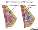
When a noncancerous tumor such as a fibroadenoma of the breast is removed, it is often called an excisional breast biopsy, instead of a lumpectomy.
Fibroadenoma
Fibroadenoma of the breast is a benign tumor. Benign tumor means it is not caused by cancer.
Description
Sometimes, the health care provider cannot feel the lump when examining you. However, it can be seen on imaging results. In this case, a wire localization will be done before the surgery.
-
A radiologist will use a
mammogram
or
ultrasound
to place a needlewire (or needlewires) in or near the abnormal breast area.
Mammogram
A mammogram is an x-ray picture of the breasts. It is used to find breast tumors and cancer.
 ImageRead Article Now Book Mark Article
Ultrasound
Ultrasound uses high-frequency sound waves to make images of organs and structures inside the body.
 ImageRead Article Now Book Mark Article
 - This will help the surgeon know where the cancer is so that it can be removed.
Breast lump removal is done as an outpatient surgery most of the time. You will be given general anesthesia (you will be asleep, but pain-free) or local anesthesia (you are awake, but sedated and pain-free). The procedure takes about 1 hour.
General anesthesia
General anesthesia is treatment with certain medicines that puts you into a deep sleep so you do not feel pain during surgery. After you receive the...
The surgeon makes a small cut on your breast. The cancer and some of the normal breast tissue around it is removed. A pathologist examines a sample of the removed tissue to make sure all the cancer has been taken out.
- When no cancer cells are found near the edges of the removed tissue, it is called a clear margin.
- Your surgeon may also remove lymph nodes in your armpit to see if the cancer has spread to them.
Sometimes, small metal clips will be placed inside the breast to mark the area of tissue removal. This makes the area easy to see on future mammograms. It also helps guide radiation therapy, when needed.
The surgeon will close your skin with stitches. These may dissolve or need to be removed later. Rarely, a drain tube may be placed to remove extra fluid. Your doctor will send the lump to a laboratory for more testing.
Why the Procedure Is Performed
Surgery to remove a breast cancer is most often the first step in treatment.
Breast cancer
Breast cancer is cancer that starts in the tissues of the breast. There are 2 main types of breast cancer:Ductal carcinoma starts in the tubes (duct...

The choice of which surgery is best for you can be difficult. It may be hard to know whether lumpectomy or mastectomy (removal of the entire breast) is best. You and the health care providers who are treating your breast cancer will decide together. In general:
-
Lumpectomy is often preferred for smaller
breast lumps
. This is because it is a smaller procedure and it has about the same chance of curing breast cancer as a
mastectomy
. It is a good option as you get to keep most of your breast.
Breast lumps
A breast lump is swelling, a growth, or a lump in the breast. Breast lumps in both men and women raise concern for breast cancer, even though most l...
 ImageRead Article Now Book Mark Article
Mastectomy
A mastectomy is surgery to remove the entire breast. Most of the time, some of the skin and the nipple are also removed. The surgery is most often ...
 ImageRead Article Now Book Mark Article
 - Mastectomy to remove all breast tissue may be done if the area of cancer is too large to remove without deforming the breast.
You and your provider should consider:
- The size of your tumor
- Where it is in your breast
- If there is more than one tumor
- How much of the breast is affected
- The size of your breasts
- Your age
- Your family history
- Your general health, including whether you have reached menopause
- If you are pregnant
Risks
Risks for any surgery are:
-
Bleeding
Bleeding
Bleeding is the loss of blood. Bleeding may be:Inside the body (internally) Outside the body (externally)Bleeding may occur:Inside the body when blo...
 ImageRead Article Now Book Mark Article
 - Infection
- Reactions to medicines
- Risks associated with general anesthesia
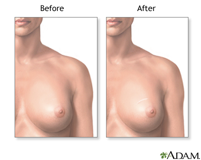
The appearance of your breast may change after surgery. You may notice dimpling, a scar, or a difference in shape between your breasts. Also, the area of the breast around the incision may be numb.
You may need another procedure to remove more breast tissue if tests show the cancer is too close to the edge of the tissue already removed.
Before the Procedure
Always tell your provider:
- If you could be pregnant
- What drugs you are taking, even drugs or herbs you bought without a prescription
During the days before your surgery:
- You may be asked to stop taking aspirin, ibuprofen (Advil, Motrin), naproxen (Aleve, Naprosyn), clopidogrel (Plavix), warfarin (Coumadin), and any other drugs that make it hard for your blood to clot.
- Ask your provider which drugs you should still take on the day of your surgery.
- If you smoke, try to stop. Your provider can help.
On the day of surgery:
- Follow your provider's instructions about eating or drinking before surgery.
- Take the drugs your provider told you to take with a small sip of water.
- Your provider will tell you when to arrive for the procedure.
After the Procedure
The recovery period is very short for a simple lumpectomy. Many women have little pain, but if you do feel pain, you can take pain medicine, such as acetaminophen.
Your skin should heal in about a month. You will need to take care of the surgical cut area . Change dressings as your provider tells you to. Watch for signs of infection when you get home (such as redness, swelling, or drainage from the incision).
Take care of the surgical cut area
Surgical incision care; Open wound care

You may need to empty a fluid drain a few times a day for 1 to 2 weeks. Your provider will remove the drain later.
Most women can go back to their usual activities in a week or so. Avoid heavy lifting, jogging, or activities that cause pain in the surgical area for 1 to 2 weeks.
Outlook (Prognosis)
The outcome of a lumpectomy for breast cancer depends mostly on the size of the cancer. It also depends on its spread to lymph nodes underneath your arm.
A lumpectomy for breast cancer is most often followed by radiation therapy and other treatments such as chemotherapy , hormone therapy , or both.
Radiation therapy
Radiation therapy uses high-powered x-rays, particles, or radioactive seeds to kill cancer cells.

Chemotherapy
The term chemotherapy is used to describe cancer-killing drugs. Chemotherapy may be used to:Cure the cancerShrink the cancerPrevent the cancer from ...

Hormone therapy
HRT- types; Estrogen replacement therapy - types; ERT- types of hormone therapy; Hormone replacement therapy - types; Menopause - types of hormone th...
You may not need a breast reconstruction after lumpectomy.
Breast reconstruction
After a mastectomy, some women choose to have cosmetic surgery to remake their breast. This type of surgery is called breast reconstruction. It can...
References
American Cancer Society. Breast-conserving surgery (lumpectomy). Updated September 29, 2016. www.cancer.org/cancer/breastcancer/detailedguide/breast-cancer-treating-breast-conserving-surgery . Accessed December 16, 2016.
Bevers TB, Brown PH, Maresso KC, Hawk ET. Cancer prevention, screening, and early detection. In: Niederhuber JE, Armitage JO, Doroshow JH, Kastan MB, Tepper JE, eds. Abeloff's Clinical Oncology. 5th ed. Philadelphia, PA: Elsevier Churchill Livingstone; 2014:chap 23.
Hunt KK, Mittendorf EA. Diseases of the breast. In: Townsend CM, Beauchamp RD, Evers BM, Mattox KL, eds. Sabiston Textbook of Surgery. 20th ed. Philadelphia, PA: Elsevier; 2017:chap 34.
The American Society of Breast Surgeons. Performance and practice guidelines for breast-conserving surgery/partial mastectomy. Updated February 22, 2015. www.breastsurgeons.org/new_layout/about/statements/PDF_Statements/PerformancePracticeGuidelines_Breast-ConservingSurgery-PartialMastectomy.pdf . Accessed December 16, 2016.
Wolff AC, Domchek SM, Davidson NE, Sacchini V, McCormick B. Cancer of the breast. In: Niederhuber JE, Armitage JO, Doroshow JH, Kastan MB, Tepper JE, eds. Abeloff's Clinical Oncology. 5th ed. Philadelphia, PA: Elsevier Churchill Livingstone; 2014:chap 91.
-
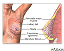
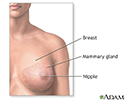
Female Breast - illustration
The female breast is either of two mammary glands (organs of milk secretion) on the chest.
Female Breast
illustration
-
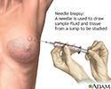
Needle biopsy of the breast - illustration
A needle biopsy is performed under local anesthesia. Simple aspirations are performed with a small gauge needle to attempt to draw fluid from lumps that are thought to be cysts. Fine needle biopsy uses a larger needle to make multiple passes through a lump, drawing out tissue and fluid. Withdrawn fluid and tissue is further evaluated to determine if there are cancerous cells present.
Needle biopsy of the breast
illustration
-
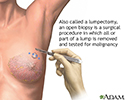
Open biopsy of the breast - illustration
An open biopsy can be performed under local or general anesthesia and will leave a small scar. Prior to surgery, a radiologist often first marks the lump with a wire, making it easier for the surgeon to find.
Open biopsy of the breast
illustration
-
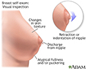
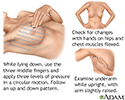
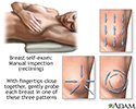
Breast self-exam - illustration
Monthly breast self-exams should always include: visual inspection (with and without a mirror) to note any changes in contour or texture; and manual inspection in standing and reclining positions to note any unusual lumps or thicknesses.
Breast self-exam
illustration
-
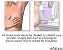
Breast lumps - illustration
Less than one-fourth of all breast lumps are found to be cancerous, but benign breast disease can be difficult to distinguish from cancer. Consequently, all breast lumps should be checked by a health care professional.
Breast lumps
illustration
-
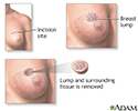
Lumpectomy - illustration
Lumpectomy is a surgical procedure performed on a solid breast mass to determine if it is malignant. The suspicious lump and some surrounding tissue is excised and analyzed.
Lumpectomy
illustration
-
Causes of breast lumps - illustration
Most breast lumps are benign (non-cancerous), as in fibroadenoma, a condition that mostly affects women under age 30. Fibrocystic breast changes occur in more than 60% of all women. Fibrocystic breast cysts change in size with the menstrual cycle, whereas a lump from fibroadenoma does not. While most breast lumps are benign, it is important to identify those that are not. See your health care provider if a lump is new, persistent, growing, hard, immobile, or causing skin deformities.
Causes of breast lumps
illustration
-
Breast lump removal - Series
Presentation
-
Breast cancer
(Alt. Medicine)
Review Date: 11/11/2016
Reviewed By: Mary C. Mancini, MD, PhD, Department of Surgery, Louisiana State University Health Sciences Center-Shreveport, Shreveport, Louisiana. Review provided by VeriMed Healthcare Network. Also reviewed by David Zieve, MD, MHA, Medical Director, Brenda Conaway, Editorial Director, and the A.D.A.M. Editorial team.
