Craniosynostosis
Premature closure of sutures; Synostosis; Plagiocephaly; Scaphocephaly; Fontanelle - craniosynostosis; Soft spot - craniosynostosis
Craniosynostosis is a birth defect in which one or more of the sutures on a baby's head closes earlier than usual.
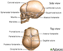
The skull of an infant or young child is made up of bony plates that allow for growth of the skull. The borders at which these plates intersect are called sutures or suture lines. The sutures between these bony plates normally close by the time the child is 2 or 3 years old.
Early closing of a suture causes the baby to have an abnormally shaped head and may limit brain growth.
Causes
The cause of craniosynostosis is unknown. Genes may play a role, but there is usually no family history of the condition.
One type that is passed down through families (inherited) can occur with other health problems, such as seizures, decreased intelligence, and blindness. Genetic disorders commonly linked to craniosynostosis include Crouzon, Apert, Carpenter, Saethre-Chotzen, and Pfeiffer syndromes.
However, most children with craniosynostosis are otherwise healthy and have normal intelligence.
Symptoms
Symptoms depend on the type of craniosynostosis. They may include:
- No "soft spot" (fontanelle) on the newborn's skull
- A raised hard ridge along the affected sutures
- Unusual head shape
- Slow or no increase in the head size over time as the baby grows
Types of craniosynostosis:
- Sagittal synostosis (scaphocephaly) is the most common type. It affects the main suture on the very top of the head. The early closing forces the head to grow long and narrow, instead of wide. Babies with this type tend to have a broad forehead. It is more common in boys than girls.
- Frontal plagiocephaly is the next most common type. It affects the suture that runs from ear to ear on the top of the head. It is more common in girls.
- Metopic synostosis is a rare form that affects the suture close to the forehead. The child's head shape may be described as trigonocephaly. It may range from mild to severe.
Exams and Tests
The health care provider will feel the infant's head and perform a physical exam.
The following tests may be done:
- Measuring the width of the infant's head
-
X-rays
of the skull
X-rays
A skull x-ray is a picture of the bones surrounding the brain, including the facial bones, the nose, and the sinuses.
 ImageRead Article Now Book Mark Article
 -
CT scan
of the head
CT scan
A head computed tomography (CT) scan uses many x-rays to create pictures of the head, including the skull, brain, eye sockets, and sinuses.
 ImageRead Article Now Book Mark Article
Well-child visits are an important part of your child's health care. They allow your provider to regularly check the growth of your infant's head over time. This will help identify any problems early.
Treatment
Surgery is done while the baby is still an infant. The goals of surgery are:
- Relieve any pressure on the brain
- Make sure there is enough room in the skull to allow the brain to properly grow
- Improve the appearance of the child's head
Outlook (Prognosis)
How well a child does depends on:
- How many sutures are involved
- The child's overall health
Children with this condition who have surgery do well in most cases, especially when the condition is not associated with a genetic syndrome.
Possible Complications
Craniosynostosis results in head deformity that can be severe and permanent if it is not corrected. Increased intracranial pressure, seizures, and developmental delay can occur.
When to Contact a Medical Professional
Call your child's provider if:
- You think your child's head shape is unusual.
- Your child is not growing well.
- The child has unusual raised ridges on the scalp.
References
Graham JM, Sanchwez-Lara PA. Craniosynostosis. In: Graham JM, Sanchez-Lara PA, eds. Smith's'Recognizable Patterns of Human Deformation . 4th ed. Philadelphia, PA: Elsevier; 2016:chap 29.
Kinsman SL, Johnston MV. Craniosynostosis. In: Kliegman RM, Stanton BF, St Geme JW, Schor NF, eds. Nelson Textbook of Pediatrics . 20th ed. Philadelphia, PA: Elsevier; 2016:chap 591.
-
Skull of a newborn - illustration
The "sutures" or anatomical lines where the bony plates of the skull join together can be easily felt in the newborn infant. The diamond shaped space on the top of the skull and the smaller space further to the back are often referred to as the "soft spot" in young infants.
Skull of a newborn
illustration
Review Date: 11/3/2015
Reviewed By: Kimberly G. Lee, MD, MSc, IBCLC, Associate Professor of Pediatrics, Division of Neonatology, Medical University of South Carolina, Charleston, SC. Review provided by VeriMed Healthcare Network. Also reviewed by David Zieve, MD, MHA, Isla Ogilvie, PhD, and the A.D.A.M. Editorial team.

