Kidney stones
Renal calculi; Nephrolithiasis; Stones - kidney; Calcium oxalate - stones; Cystine - stones; Struvite - stones; Uric acid - stones; Urinary lithiasis
A kidney stone is a solid mass made up of tiny crystals. One or more stones can be in the kidney or ureter at the same time.
Causes
Cystinuria is a rare condition in which stones made from an amino acid called cysteine form in the kidney, ureter, and bladder. Cystine is formed wh...

Kidney stones are common. Some types run in families. They often occur in premature infants.
There are different types of kidney stones. The cause of the problem depends on the type of stone.
Stones can form when urine contains too much of certain substances that form crystals. These crystals can develop into stones over weeks or months.
- Calcium stones are most common. They are most likely to occur in men between ages 20 to 30. Calcium can combine with other substances to form the stone.
- Oxalate is the most common of these. Oxalate is present in certain foods such as spinach. It is also found in vitamin C supplements. Diseases of the small intestine increase your risk of these stones.
Calcium stones can also form from combining with phosphate or carbonate.
Other types of stones include:
-
Cystine stones can form in people who have
cystinuria
. This disorder runs in families. It affects both men and women.
Cystinuria
Cystinuria is a rare condition in which stones made from an amino acid called cysteine form in the kidney, ureter, and bladder. Cystine is formed wh...
 ImageRead Article Now Book Mark Article
 - Struvite stones are mostly found in women who have a urinary tract infection. These stones can grow very large and can block the kidney, ureter, or bladder.
- Uric acid stones are more common in men than in women. They can occur with gout or chemotherapy.
- Other substances such as certain medicines also can form stones.
The biggest risk factor for kidney stones is not drinking enough fluids. Kidney stones are more likely to occur if you make less than 1 liter (32 ounces) of urine a day.
Symptoms
You may not have symptoms until the stones move down the tubes (ureters) through which urine empties into your bladder. When this happens, the stones can block the flow of urine out of the kidneys.
The main symptom is severe pain that starts and stops suddenly:
- Pain may be felt in the belly area or side of the back.
-
Pain may move to groin area (
groin pain
) or testicles (
testicle pain
).
Groin pain
Groin pain refers to discomfort in the area where the abdomen ends and the legs begin. This article focuses on groin pain in men. The terms "groin"...
Read Article Now Book Mark ArticleTesticle pain
Testicle pain is discomfort in one or both testicles. The pain can spread into the lower abdomen.
 ImageRead Article Now Book Mark Article
Other symptoms can include:
-
Abnormal urine color
Abnormal urine color
The usual color of urine is straw-yellow. Abnormally colored urine may be cloudy, dark, or blood-colored.
 ImageRead Article Now Book Mark Article
 -
Blood in the urine
Blood in the urine
Blood in your urine is called hematuria. The amount may be very small and only detected with urine tests or under a microscope. In other cases, the...
 ImageRead Article Now Book Mark Article
 - Chills
-
Fever
Fever
Fever is the temporary increase in the body's temperature in response to a disease or illness. A child has a fever when the temperature is at or abov...
 ImageRead Article Now Book Mark Article
 -
Nausea and vomiting
Nausea and vomiting
Nausea is feeling an urge to vomit. It is often called "being sick to your stomach. "Vomiting or throwing-up is forcing the contents of the stomach ...
 ImageRead Article Now Book Mark Article
Exams and Tests
The health care provider will perform a physical exam. The belly area (abdomen) or back might feel sore.
Tests that may be done include:
- Blood tests to check calcium, phosphorus, uric acid, and electrolyte levels
- Kidney function tests
-
Urinalysis
to see crystals and look for
red blood cells in urine
Urinalysis
Urinalysis is the physical, chemical, and microscopic examination of urine. It involves a number of tests to detect and measure various compounds th...
 ImageRead Article Now Book Mark Article
Red blood cells in urine
The RBC urine test measures the number of red blood cells in a urine sample.
 ImageRead Article Now Book Mark Article
 - Examination of the stone to determine the type
Stones or a blockage can be seen on:
-
Abdominal CT scan
Abdominal CT scan
An abdominal CT scan is an imaging method. This test uses x-rays to create cross-sectional pictures of the belly area. CT stands for computed tomog...
 ImageRead Article Now Book Mark Article
 -
Abdominal/kidney MRI
Abdominal/kidney MRI
An abdominal magnetic resonance imaging scan is an imaging test that uses powerful magnets and radio waves. The waves create pictures of the inside ...
 ImageRead Article Now Book Mark Article
 -
Abdominal x-rays
Abdominal x-rays
An abdominal x-ray is an imaging test to look at organs and structures in the abdomen. Organs include the spleen, stomach, and intestines. When the ...
 ImageRead Article Now Book Mark Article
 -
Intravenous pyelogram
(
IVP
)
Intravenous pyelogram
An intravenous pyelogram (IVP) is a special x-ray exam of the kidneys, bladder, and ureters (the tubes that carry urine from the kidneys to the bladd...
 ImageRead Article Now Book Mark Article
IVP
An intravenous pyelogram (IVP) is a special x-ray exam of the kidneys, bladder, and ureters (the tubes that carry urine from the kidneys to the bladd...
 ImageRead Article Now Book Mark Article
 -
Kidney ultrasound
Kidney ultrasound
Abdominal ultrasound is a type of imaging test. It is used to look at organs in the abdomen, including the liver, gallbladder, spleen, pancreas, and...
 ImageRead Article Now Book Mark Article
 - Retrograde pyelogram
Treatment
Treatment depends on the type of stone and the severity of your symptoms.
Kidney stones that are small most often pass through your system on their own.
- Your urine should be strained so the stone can be saved and tested.
-
Drink at least 6 to 8 glasses of water per day to produce a large amount of urine. This will help the
stone pass
.
Stone pass
Renal calculi and self-care; Nephrolithiasis and self-care; Stones and kidney - self-care; Calcium stones and self-care; Oxalate stones and self-care...
 ImageRead Article Now Book Mark Article
 - Pain can be very bad. Over-the-counter pain medicines (for example, ibuprofen and naproxen), either alone or along with narcotics, can be very effective.
Some people with severe pain from kidney stones need to stay in the hospital. You may need to get fluids through a vein.
For some types of stones, your provider may prescribe medicine to prevent stones from forming or help break down and remove the material that is causing the stone. These medicines can include:
-
Allopurinol (for
uric acid
stones)
Uric acid
Uric acid is a chemical created when the body breaks down substances called purines. Purines are found in some foods and drinks. These include live...
 ImageRead Article Now Book Mark Article
 - Antibiotics (for struvite stones)
- Diuretics (water pills)
- Phosphate solutions
- Sodium bicarbonate or sodium citrate
- Water pills (thiazide diuretics)
- Tamsulosin to relax the ureter and help the stone pass
Surgery is often needed if:
- The stone is too large to pass on its own
- The stone is growing
- The stone is blocking urine flow and causing an infection or kidney damage
- The pain cannot be controlled
Today, most treatments are much less invasive than in the past.
-
Lithotripsy
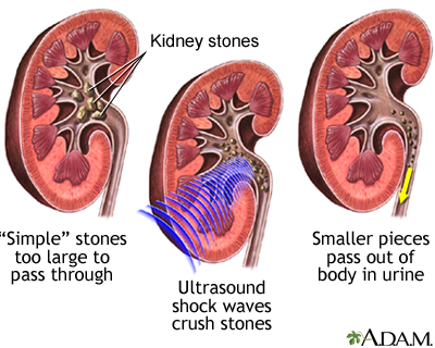
is used to remove stones slightly smaller than a half an inch (1.25 centimeters) that are located in the kidney or ureter. It uses sound or shock waves to break up stones. Then, the stone fragments leave the body in the urine. It is also called extracorporeal shock-wave lithotripsy or ESWL.
Lithotripsy
Lithotripsy is a procedure that uses shock waves to break up stones in the kidney, bladder, or ureter (tube that carries urine from your kidneys to y...
 ImageRead Article Now Book Mark Article
 -
Procedures performed by
passing a special instrument
through a small surgical cut in your skin and into your kidney or ureters are used for large stones, or when the kidneys or surrounding areas are incorrectly formed. The stone is removed with a tube (endoscope).
Passing a special instrument
Percutaneous (through the skin) urinary procedures help drain urine from your bladder and get rid of kidney stones.
Read Article Now Book Mark Article - Ureteroscopy may be used for stones in the lower urinary tract.
- Rarely, open surgery (nephrolithotomy) may be needed if other methods do not work or are not possible.
Talk to your provider about what treatment options may work for you.
Talk to your provider
Nephrolithiasis - what to ask your doctor; Renal calculi - what to ask your doctor; What to ask your doctor about kidney stones
You will need to take self-care steps. Which steps you take depend on the type of stone you have, but they may include:
Self-care steps.
Renal calculi and self-care; Nephrolithiasis and self-care; Stones and kidney - self-care; Calcium stones and self-care; Oxalate stones and self-care...

- Drinking extra water and other liquids
- Eating more of some foods and cutting back on other foods
- Taking medicines to help prevent stones
- Taking medicines to help you pass a stone (anti-inflammatory drugs, alpha-blockers)
Outlook (Prognosis)
Kidney stones are painful but most of the time can be removed from the body without causing lasting damage.
Kidney stones often come back. This occurs more often if the cause is not found and treated.
You are at risk for:
- Urinary tract infection
- Kidney damage or scarring if treatment is delayed for too long
Possible Complications
Complication of kidney stones may include the obstruction of the ureter (acute unilateral obstructive uropathy).
When to Contact a Medical Professional
Call your provider if you have symptoms of a kidney stone:
- Severe pain in your back or side that will not go away
- Blood in your urine
- Fever and chills
- Vomiting
- Urine that smells bad or looks cloudy
- A burning feeling when you urinate
If you have been diagnosed with blockage from a stone, passage must be confirmed either by capture in a strainer during urination or by follow-up x-ray.
Prevention
If you have a history of stones:
- Drink plenty of fluids (6 to 8 glasses of water per day) to produce enough urine.
- You may need to take medicine or make changes to your diet for some types of stones.
- Your provider may want to do blood and urine tests to help determine the proper prevention steps.
References
Bushinsky DA. Nephrolithiasis. In: Goldman L, Schafer AI, eds. Goldman's Cecil Medicine . 25th ed. Philadelphia, PA: Elsevier Saunders; 2016:chap 126.
Fink HA, Wilt TJ, Eidman KE, et al. Medical management to prevent recurrent nephrolithiasis in adults: a systematic review for an American College of Physicians Clinical Guideline. Ann Intern Med . 2013;158(7):535-543. PMID: 23546565 www.ncbi.nlm.nih.gov/pubmed/23546565 .
Fink HA, Wilt TJ, Eidman KE, et al. Recurrent nephrolithiasis in adults: comparative effectiveness of preventive medical strategies [Internet]. Rockville, MD. Agency for Healthcare Research and Quality (US) 2012 ; Report No.: 12-EHC049-EF. PMID: 22896859 www.ncbi.nlm.nih.gov/pubmed/22896859 .
Lipkin ME, Ferrandino MN, Preminger Glenn M. Evaluation and medical management of urinary lithiasis. In: Wein AJ, Kavoussi LR, Partin AW, Peters CA, eds. Campbell-Walsh Urology . 11th ed. Philadelphia, PA: Elsevier; 2016:chap 52.
Qaseem A, Dallas P, Forciea MA, Starkey M, Denberg TD; Clinical Guidelines Committee of the American College of Physicians. Dietary and pharmacologic management to prevent recurrent nephrolithiasis in adults: a clinical practice guideline from the American College of Physicians. Ann Intern Med . 2014;161(9):659-667. PMID: 25364887 www.ncbi.nlm.nih.gov/pubmed/25364887 .
-
Kidney stones
Animation
-
Kidney stones
Animation
-
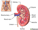
Kidney anatomy - illustration
The kidneys are responsible for removing wastes from the body, regulating electrolyte balance and blood pressure, and stimulating red blood cell production.
Kidney anatomy
illustration
-
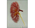
Kidney - blood and urine flow - illustration
This is the typical appearance of the blood vessels (vasculature) and urine flow pattern in the kidney. The blood vessels are shown in red and the urine flow pattern in yellow.
Kidney - blood and urine flow
illustration
-
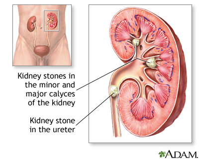
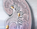
Nephrolithiasis - illustration
Kidney stones result when urine becomes too concentrated and substances in the urine crystalize to form stones. Symptoms arise when the stones begin to move down the ureter causing intense pain. Kidney stones may form in the pelvis or calyces of the kidney or in the ureter.
Nephrolithiasis
illustration
-
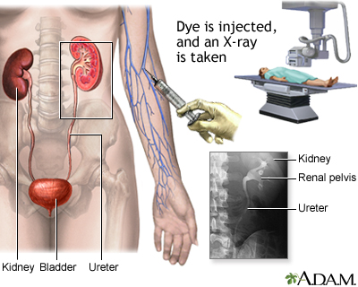
Intravenous pyelogram (IVP) - illustration
In the procedure intravenous pyelogram (IVP), the patient is injected with radiopaque dye and X-rays are taken as the dye travels through the urinary tract. This procedure is performed to confirm the presence of kidney stones, although some stones may be too small to see.
Intravenous pyelogram (IVP)
illustration
-
Lithotripsy procedure - illustration
Extracorporeal shock wave lithotripsy (ESWL) is a procedure used to shatter simple stones in the kidney or upper urinary tract. Ultrasonic waves are passed through the body until they strike the dense stones. Pulses of sonic waves pulverize the stones, which are then more easily passed through the ureter and out of the body in the urine.
Lithotripsy procedure
illustration
-
Kidney stones
(In-Depth)
-
Kidney stones
(Alt. Medicine)
Review Date: 8/31/2015
Reviewed By: Jennifer Sobol, DO, urologist at the Michigan Institute of Urology, West Bloomfield, MI. Review provided by VeriMed Healthcare Network. Internal review and update on 09/01/2016 by David Zieve, MD, MHA, Isla Ogilvie, PhD, and the A.D.A.M. Editorial team.

