Episcleritis
Episcleritis is irritation and inflammation of the episclera, a thin layer of tissue covering the white part (sclera) of the eye. It is not an infection.
Causes
Episcleritis is a common condition. In most cases the problem is mild and vision is normal.
The cause is often unknown. But, it may occur with certain diseases, such as:
-
Herpes zoster
Herpes zoster
Shingles (herpes zoster) is a painful, blistering skin rash. It is caused by the varicella-zoster virus. This is the virus that also causes chicken...
 ImageRead Article Now Book Mark Article
 -
Rheumatoid arthritis
Rheumatoid arthritis
Rheumatoid arthritis (RA) is a long-term disease. It leads to inflammation of the joints and surrounding tissues. It can also affect other organs....
 ImageRead Article Now Book Mark Article
 -
Sjogren syndrome
Sjogren syndrome
Sjögren syndrome is an autoimmune disorder in which the glands that produce tears and saliva are destroyed. This causes dry mouth and dry eyes. The...
 ImageRead Article Now Book Mark Article
 - Syphilis
-
Tuberculosis
Tuberculosis
Pulmonary tuberculosis (TB) is a contagious bacterial infection that involves the lungs. It may spread to other organs.
 ImageRead Article Now Book Mark Article
Symptoms
Symptoms include:
- A pink or purple color to the normally white part of the eye
-
Eye pain
Eye pain
Pain in the eye may be described as a burning, throbbing, aching, or stabbing sensation in or around the eye. It may also feel like you have a forei...
Read Article Now Book Mark Article - Eye tenderness
-
Sensitivity to light
Sensitivity to light
Photophobia is eye discomfort in bright light.
 ImageRead Article Now Book Mark Article
 -
Tearing of the eye
Tearing of the eye
Watery eyes means you have too many tears draining from the eyes. Tears help keep the surface of the eye moist. They wash away particles and foreig...
ImageRead Article Now Book Mark Article
Exams and Tests
Your health care provider will do an eye exam to diagnose the disorder. Most of the time, no special tests are needed.
Treatment
The condition most often goes away on its own in 1 to 2 weeks. Using corticosteroid eye drops may help ease the symptoms faster.
Outlook (Prognosis)
Episcleritis most often improves without treatment. However, treatment may make symptoms go away sooner.
Possible Complications
In some cases, the condition may return. Rarely, irritation and inflammation of the white part of the eye may develop. This is called scleritis.
When to Contact a Medical Professional
Call your health care provider if you have symptoms of episcleritis that last for more than 2 weeks. Get checked again if your pain gets worse or you have problems with your vision.
References
Goldstein DA, Patel SS, Tessler HH. Episcleritis and scleritis. In: Yanoff M, Duker JS, eds. Ophthalmology . 4th ed. St Louis, MO: Mosby Elsevier; 2014:chap 4.11.
Watson P. Diseases of the sclera and episclera. In: Tasman W, Jaeger EA, eds. Duane's Ophthalmology. 2013 ed. Philadelphia, PA: Lippincott Williams & Wilkins; 2013:vol 4;chap 23.
Yanoff M, Cameron JD. Diseases of the visual system. In: Goldman L, Schafer AI, eds. Goldman's Cecil Medicine . 25th ed. Philadelphia, PA: Elsevier Saunders; 2016:chap 423.
-
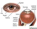
External and internal eye anatomy - illustration
The cornea allows light to enter the eye. As light passes through the eye the iris changes shape by expanding and letting more light through or constricting and letting less light through to change pupil size. The lens then changes shape to allow the accurate focusing of light on the retina. Light excites photoreceptors that eventually, through a chemical process, transmit nerve signals through the optic nerve to the brain. The brain processes these nerve impulses into sight.
External and internal eye anatomy
illustration
-
External and internal eye anatomy - illustration
The cornea allows light to enter the eye. As light passes through the eye the iris changes shape by expanding and letting more light through or constricting and letting less light through to change pupil size. The lens then changes shape to allow the accurate focusing of light on the retina. Light excites photoreceptors that eventually, through a chemical process, transmit nerve signals through the optic nerve to the brain. The brain processes these nerve impulses into sight.
External and internal eye anatomy
illustration
Review Date: 8/20/2016
Reviewed By: Franklin W. Lusby, MD, ophthalmologist, Lusby Vision Institute, La Jolla, CA. Also reviewed by David Zieve, MD, MHA, Isla Ogilvie, PhD, and the A.D.A.M. Editorial team.