Culture - colonic tissue
Colonic tissue culture
A colonic tissue culture is a lab test to check for the cause of disease. The sample of tissue for the test is taken from the large intestine. The cause may be bacteria, fungi, or viruses.
How the Test is Performed
The health care provider removes a piece of tissue from your large intestine. This is done during a colonoscopy .
Colonoscopy
A colonoscopy is an exam that views the inside of the colon (large intestine) and rectum, using a tool called a colonoscope. The colonoscope has a sm...

- The sample is sent to a lab.
- It is placed in a special dish that contains a gel. Bacteria and other organisms can grow in this gel. The dish is then stored at a certain temperature.
- The lab team checks the sample daily. They check to see if bacteria, viruses, or fungi have grown.
If certain germs grow, more tests will be done to identify them. This helps decide the best treatment.
How to Prepare for the Test
There is no specific preparation needed for a culture.
How the Test will Feel
Once the sample is taken, the culture does not involve you. Therefore, there is no pain.
Why the Test is Performed
Your provider may order this test if you have signs or symptoms of a large intestine infection. A culture is often done when other tests such as a stool culture could not identify the cause of infection.
Stool culture
A fecal culture is a lab test to find organisms in the stool (feces) that can cause gastrointestinal symptoms and disease.

Normal Results
A normal result means that no disease-causing organisms have grown in the lab dish.
Some "healthy" bacteria, called bowel flora, are normally found in the gut. The growth of such bacteria during this test does not mean there is an infection.
Normal value ranges may vary slightly among different laboratories. Talk to your provider about your test results.
What Abnormal Results Mean
An abnormal result means that disease-causing organisms have grown in the lab dish. These organisms may include:
- Clostridium difficile bacteria
- Cytomegalovirus
- Mycobacterium tuberculosis bacteria
-
Salmonella bacteria
Salmonella bacteria
Salmonella enterocolitis is an infection in the lining of the small intestine that is caused by salmonella bacteria. It is a type of food poisoning....
ImageRead Article Now Book Mark Article - Shigella bacteria
These organisms may lead to diarrhea or colon infections.

Risks
There are no risks in a colonic tissue culture.
References
Beavis KG, Charnot-Katsikas A. Specimen collection and handling for diagnosis of infectious diseases. In: McPherson RA, Pincus MR, eds. Henry's Clinical Diagnosis and Management by Laboratory Methods . 23rd ed. Philadelphia, PA: Elsevier; 2017:chap 64.
DuPont HL. Approach to the patient with suspected enteric infection. In: Goldman L, Schafer AI, eds. Goldman's Cecil Medicine . 25th ed. Philadelphia, PA: Elsevier Saunders; 2016:chap 283.
Hall GS, Woods GL. Medical bacteriology. In: McPherson RA, Pincus MR, eds. Henry's Clinical Diagnosis and Management by Laboratory Methods . 23rd ed. Philadelphia, PA: Elsevier; 2017:chap 58.
Haines CF, Sears CL. Infectious enteritis and proctocolitis. In: Feldman M, Friedman LS, Brandt LJ, eds. Sleisenger and Fordtran's Gastrointestinal and Liver Disease . 10th ed. Philadelphia, PA: Elsevier Saunders; 2016:chap 110.
Semrad CE. Approach to the patient with diarrhea and malabsorption. In: Goldman L, Schafer AI, eds. Goldman's Cecil Medicine . 25th ed. Philadelphia, PA: Elsevier Saunders; 2016:chap 140.
Siddiqi HA, Salwen MJ, Shaikh MF, Bowne WB. Laboratory diagnosis of gastrointestinal and pancreatic disorders. In: McPherson RA, Pincus MR, eds. Henry's Clinical Diagnosis and Management by Laboratory Methods . 23rd ed. Philadelphia, PA: Elsevier; 2017:chap 22.
-
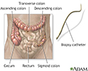
Colonoscopy - illustration
A sample of colon tissue is obtained during a colonoscopy procedure using special instruments. The specimen is sent to the laboratory and fixed with special stains. The specimen is examined under the microscope for abnormal findings, such as cancer or inflammation.
Colonoscopy
illustration
-
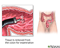
Colon culture - illustration
When polyps are discovered in a sigmoidoscopy (an inspection of the lower third of the large intestine), they are retrieved to be tested for cancer. If a large amount of polyps are found, a more thorough examination of the entire length of the large intestine (a colonoscopy) may be recommended.
Colon culture
illustration
-
Colonoscopy - illustration
A sample of colon tissue is obtained during a colonoscopy procedure using special instruments. The specimen is sent to the laboratory and fixed with special stains. The specimen is examined under the microscope for abnormal findings, such as cancer or inflammation.
Colonoscopy
illustration
-
Colon culture - illustration
When polyps are discovered in a sigmoidoscopy (an inspection of the lower third of the large intestine), they are retrieved to be tested for cancer. If a large amount of polyps are found, a more thorough examination of the entire length of the large intestine (a colonoscopy) may be recommended.
Colon culture
illustration
Review Date: 5/11/2016
Reviewed By: Subodh K. Lal, MD, gastroenterologist with Gastrointestinal Specialists of Georgia, Austell, GA. Review provided by VeriMed Healthcare Network. Also reviewed by David Zieve, MD, MHA, Isla Ogilvie, PhD, and the A.D.A.M. Editorial team.