Heart pacemaker
Cardiac pacemaker implantation; Artificial pacemaker; Permanent pacemaker; Internal pacemaker; Cardiac resynchronization therapy; CRT; Biventricular pacemaker; Arrhythmia - pacemaker; Abnormal heart rhythm - pacemaker; Bradycardia - pacemaker; Heart block - pacemaker; Mobitz - pacemaker; Heart failure - pacemaker; HF - pacemaker; CHF- pacemaker
A pacemaker is a small, battery-operated device. This device senses when your heart is beating irregularly or too slowly . It sends a signal to your heart that makes your heart beat at the correct pace.
Irregularly or too slowly
Palpitations are feelings or sensations that your heart is pounding or racing. They can be felt in your chest, throat, or neck. You may:Have an unpl...


Description
Newer pacemakers weigh as little as 1 ounce (28 grams). Most pacemakers have 2 parts:
- The generator contains the battery and the information to control the heartbeat.
- The leads are wires that connect the heart to the generator and carry the electrical messages to the heart.
A pacemaker must be implanted under the skin. This procedure takes about 1 hour in most cases. You will be given a sedative to help you relax. You will be awake during the procedure.
A small incision (cut) is made. Most often, the cut is on the left side of the chest below your collarbone. The pacemaker generator is then placed under the skin at this location. The generator may also be placed in the abdomen, but this is less common.
Using live x-rays to see the area, the doctor puts the leads through the cut, into a vein, and then into the heart. The leads are connected to the generator. The skin is closed with stitches. Most people go home within 1 day of the procedure.
There are 2 kinds of pacemakers used only in medical emergencies. They are:
- Transcutaneous pacemakers
- Transvenous pacemakers
They are not permanent pacemakers.
Why the Procedure Is Performed
Pacemakers may be used for people who have heart problems that cause their heart to beat too slowly. A slow heartbeat is called bradycardia . Two common problems that cause a slow heartbeat are sinus node disease and heart block.
Bradycardia
An arrhythmia is a disorder of the heart rate (pulse) or heart rhythm. The heart can beat too fast (tachycardia), too slow (bradycardia), or irregul...

Sinus node disease
Normally, the heartbeat starts in an area in the top chambers of the heart (atria). This area is the heart's pacemaker. It may be called the sinoat...

When your heart beats too slowly, your body and brain may not get enough oxygen. Symptoms may be
-
Lightheadedness
Lightheadedness
Dizziness is a term that is often used to describe 2 different symptoms: lightheadedness and vertigo. Lightheadedness is a feeling that you might fai...
 ImageRead Article Now Book Mark Article
 -
Tiredness
Tiredness
Fatigue is a feeling of weariness, tiredness, or lack of energy.
Read Article Now Book Mark Article -
Fainting spells
Fainting spells
Fainting is a brief loss of consciousness due to a drop in blood flow to the brain. The episode most often lasts less than a couple of minutes and y...
Read Article Now Book Mark Article -
Shortness of breath
Shortness of breath
Breathing difficulty may involve:Difficult breathingUncomfortable breathingFeeling like you are not getting enough air
 ImageRead Article Now Book Mark Article
Some pacemakers can be used to stop a heart rate that is too fast ( tachycardia ) or that is irregular.
Tachycardia
An arrhythmia is a disorder of the heart rate (pulse) or heart rhythm. The heart can beat too fast (tachycardia), too slow (bradycardia), or irregul...
Other types of pacemakers can be used in severe heart failure . These are called biventricular pacemakers. They help coordinate the beating of the heart chambers.
Heart failure
Heart failure is a condition in which the heart is no longer able to pump oxygen-rich blood to the rest of the body efficiently. This causes symptom...
Most biventricular pacemakers implanted today can also work as implantable cardioverter defibrillators (ICD). ICD restore a normal heartbeat by delivering a larger shock when a potentially deadly fast heart rhythm occurs.
Implantable cardioverter defibrillators
An implantable cardioverter-defibrillator (ICD) is a device that detects any life-threatening, rapid heartbeat. This abnormal heartbeat is called an...

Risks
Possible complications of pacemaker surgery are:
-
Abnormal heart rhythms
Abnormal heart rhythms
Palpitations are feelings or sensations that your heart is pounding or racing. They can be felt in your chest, throat, or neck. You may:Have an unpl...
ImageRead Article Now Book Mark Article -
Bleeding
Bleeding
Bleeding is the loss of blood. Bleeding may be:Inside the body (internally) Outside the body (externally)Bleeding may occur:Inside the body when blo...
 ImageRead Article Now Book Mark Article
 - Punctured lung. This is rare.
- Infection
- Puncture of the heart, which can lead to bleeding around the heart. This is rare.
A pacemaker senses if the heartbeat is above a certain rate. When it is above that rate, the pacemaker will stop sending signals to the heart. The pacemaker can also sense when the heartbeat slows down too much. It will automatically turn back on and start pacing the heart again.
Before the Procedure
Always tell your health care provider about all the drugs you are taking, even drugs or herbs you bought without a prescription.
The day before your surgery:
- Shower and shampoo well.
- You may be asked to wash your whole body below your neck with a special soap.
On the day of the surgery:
- You may be asked not to drink or eat anything after midnight the night before your procedure. This includes chewing gum and breath mints. Rinse your mouth with water if it feels dry, but be careful not to swallow.
- Take the drugs you have been told to take with a small sip of water.
Your provider will tell you when to arrive at the hospital.
After the Procedure
You will probably be able to go home after 1 day. You should be able to return to your normal activity level quickly.
Ask your provider how much you can use the arm on the side of your body where the pacemaker was placed. You may be advised not to:
You can use the arm
Cardiac pacemaker implantation - discharge; Artificial pacemaker - discharge; Permanent pacemaker - discharge; Internal pacemaker - discharge; Cardia...
- Lift anything heavier than 10 to 15 pounds (4.5 to 6.75 kilograms)
- Push, pull, and twist your arm for 2 to 3 weeks.
- Raise your arm above your shoulder for several weeks.
When you leave the hospital, you will be given a card to keep in your wallet. This card lists the details of your pacemaker and has contact information for emergencies. You should always carry this wallet card with you. You should try to remember the name of the pacemaker manufacturer if you can in case you lose your card.
Outlook (Prognosis)
Pacemakers can help keep your heart rhythm and heart rate at a safe level for you. The pacemaker battery lasts about 6 to 15 years. Your provider will check the battery regularly and replace it when necessary.
References
Epstein AE, DiMarco JP, Ellenbogen KA, et al. 2012 ACCF/AHA/HRS focused update incorporated into the ACCF/AHA/HRS 2008 guidelines for device-based therapy of cardiac rhythm abnormalities: a report of the American College of Cardiology Foundation/American Heart Association Task Force on practice guidelines and the Heart Rhythm Society. J Am Coll Cardiol . 2013;61(3):e6-e75. PMID: 23265327 www.ncbi.nlm.nih.gov/pubmed/23265327 .
Olgin JE, Zipes DP. Specific arrhythmias. In: Mann DL, Zipes DP, Libby P, Bonow RO, Braunwald E, eds. Braunwald's Heart Disease: A Textbook of Cardiovascular Medicine . 10th ed. Philadelphia, PA: Elsevier Saunders; 2015:chap 37.
Swerdlow CD, Wang PJ, Zipes DP. Pacemakers and implantable cardioverter-defibrillators. In: Mann DL, Zipes DP, Libby P, Bonow RO, Braunwald E, eds. Braunwald's Heart Disease: A Textbook of Cardiovascular Medicine . 10th ed. Philadelphia, PA: Elsevier Saunders; 2015:chap 36.
-
Heartbeat
Animation
-
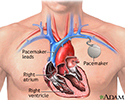
Pacemaker - illustration
A pacemaker is a small, battery-operated electronic device which is inserted under the skin to help the heart beat regularly and at an appropriate rate. The pacemaker has leads that travel through a large vein to the heart, where the wires are anchored. The leads send the electrical impulses to the heart to tell it to beat.
Pacemaker
illustration
-
Pacemaker - illustration
A pacemaker is a small, battery-operated electronic device which is inserted under the skin to help the heart beat regularly and at an appropriate rate. The pacemaker has leads that travel through a large vein to the heart, where the wires are anchored. The leads send the electrical impulses to the heart to tell it to beat.
Pacemaker
illustration
Review Date: 8/2/2016
Reviewed By: Michael A. Chen, MD, PhD, Associate Professor of Medicine, Division of Cardiology, Harborview Medical Center, University of Washington Medical School, Seattle, WA. Also reviewed by David Zieve, MD, MHA, Isla Ogilvie, PhD, and the A.D.A.M. Editorial team.