Cerebral hypoxia
Hypoxic encephalopathy; Anoxic encephalopathy
Cerebral hypoxia occurs when there is not enough oxygen getting to the brain. The brain needs a constant supply of oxygen and nutrients to function.
Cerebral hypoxia affects the largest parts of the brain, called the cerebral hemispheres. However, the term is often used to refer to a lack of oxygen supply to the entire brain.
Causes
In cerebral hypoxia, sometimes only the oxygen supply is interrupted. This can be caused by:
- Breathing in smoke (smoke inhalation), such as during a fire
-
Carbon monoxide poisoning
Carbon monoxide poisoning
Carbon monoxide is an odorless gas that causes thousands of deaths each year in North America. Breathing in carbon monoxide is very dangerous. It i...
Read Article Now Book Mark Article - Choking
-
Diseases that prevent movement (paralysis) of the breathing muscles, such as
amyotrophic lateral sclerosis
(ALS)
Amyotrophic lateral sclerosis
Amyotrophic lateral sclerosis, or ALS, is a disease of the nerve cells in the brain, brain stem and spinal cord that control voluntary muscle movemen...
 ImageRead Article Now Book Mark Article
 - High altitudes
- Pressure on (compression) the windpipe (trachea)
- Strangulation
In other cases, both oxygen and nutrient supply are stopped, caused by:
- Cardiac arrest (when the heart stops pumping)
-
Cardiac
arrhythmia
(heart rhythm problems)
Arrhythmia
An arrhythmia is a disorder of the heart rate (pulse) or heart rhythm. The heart can beat too fast (tachycardia), too slow (bradycardia), or irregul...
 ImageRead Article Now Book Mark Article
 -
Complications of
general anesthesia
General anesthesia
General anesthesia is treatment with certain medicines that puts you into a deep sleep so you do not feel pain during surgery. After you receive the...
Read Article Now Book Mark Article - Drowning
- Drug overdose
-
Injuries to a newborn that occurred before, during, or soon after birth such as
cerebral palsy
Cerebral palsy
Cerebral palsy is a group of disorders that can involve brain and nervous system functions, such as movement, learning, hearing, seeing, and thinking...
 ImageRead Article Now Book Mark Article
 -
Stroke
Stroke
A stroke occurs when blood flow to a part of the brain stops. A stroke is sometimes called a "brain attack. " If blood flow is cut off for longer th...
 ImageRead Article Now Book Mark Article
 -
Very
low blood pressure
Low blood pressure
Low blood pressure occurs when blood pressure is much lower than normal. This means the heart, brain, and other parts of the body do not get enough ...
Read Article Now Book Mark Article
Brain cells are very sensitive to a lack of oxygen. Some brain cells start dying less than 5 minutes after their oxygen supply disappears. As a result, brain hypoxia can rapidly cause severe brain damage or death.
Symptoms
Symptoms of mild cerebral hypoxia include:
- Change in attention (inattentiveness)
- Poor judgment
- Uncoordinated movement
Symptoms of severe cerebral hypoxia include:
- Complete unawareness and unresponsiveness (coma)
- No breathing
- No response of the pupils of the eye to light
Exams and Tests
Cerebral hypoxia can usually be diagnosed based on the person's medical history and a physical exam. Tests are done to determine the cause of the hypoxia, and may include:
- Angiogram of the brain
-
Blood tests, including
arterial blood gases
and blood chemical levels
Arterial blood gases
Blood gases are a measurement of how much oxygen and carbon dioxide are in your blood. They also determine the acidity (pH) of your blood.
 ImageRead Article Now Book Mark Article
 -
CT scan of the head
CT scan of the head
A head computed tomography (CT) scan uses many x-rays to create pictures of the head, including the skull, brain, eye sockets, and sinuses.
 ImageRead Article Now Book Mark Article
 -
Echocardiogram
which uses ultrasound to view the heart
Echocardiogram
An echocardiogram is a test that uses sound waves to create pictures of the heart. The picture and information it produces is more detailed than a s...
 ImageRead Article Now Book Mark Article
 -
Electrocardiogram
(ECG), a measurement of the heart's electrical activity
Electrocardiogram
An electrocardiogram (ECG) is a test that records the electrical activity of the heart.
 ImageRead Article Now Book Mark Article
 -
Electroencephalogram
(EEG), a test of brain waves that can identify seizures and show how well brain cells work
Electroencephalogram
An electroencephalogram is a test to measure the electrical activity of the brain.
 ImageRead Article Now Book Mark Article
 -
Evoked potentials
, a test that determines whether certain sensations, such as vision and touch, reach the brain
Evoked potentials
Brainstem auditory evoked response (BAER) is a test to measure the brain wave activity that occurs in response to clicks or certain tones.
 ImageRead Article Now Book Mark Article
 -
Magnetic resonance imaging
(MRI) of the head
Magnetic resonance imaging
A magnetic resonance imaging (MRI) scan is an imaging test that uses powerful magnets and radio waves to create pictures of the body. It does not us...
 ImageRead Article Now Book Mark Article
If only blood pressure and heart function remain, the brain may be completely dead.
Treatment
Cerebral hypoxia is an emergency condition that needs to be treated right away. The sooner the oxygen supply is restored to the brain, the lower the risk for severe brain damage and death.
Treatment depends on the cause of the hypoxia. Basic life support is most important. Treatment involves:
- Breathing assistance (mechanical ventilation) and oxygen
- Controlling the heart rate and rhythm
- Fluids, blood products, or medicines to raise blood pressure if it is low
- Medicines or general anesthetics to calm seizures
Sometimes a person with cerebral hypoxia is cooled to slow down the activity of the brain cells and decrease their need for oxygen. However, the benefit of this treatment has not been firmly established.
Outlook (Prognosis)
The outlook depends on the extent of the brain injury. This is determined by how long the brain lacked oxygen, and whether nutrition to the brain was also affected.
If the brain lacked oxygen for only a brief period, a coma may be reversible and the person may have a full or partial return of function. Some people recover many functions, but have abnormal movements, such as twitching or jerking, called myoclonus. Seizures may sometimes occur, and may be continuous (status epilepticus).
Most people who make a full recovery were only briefly unconscious. The longer a person is unconscious, the higher the risk for death or brain death, and the lower the chances of recovery.
Possible Complications
Complications of cerebral hypoxia include a prolonged vegetative state. This means the person may have basic life functions, such as breathing, blood pressure, sleep-wake cycle, and eye opening, but the person is not alert and does not respond to their surroundings. Such people usually die within a year, although some may survive longer.
Length of survival depends partly on how much care is taken to prevent other problems. Major complications may include:
- Bed sores
-
Clots in the veins (
deep vein thrombosis
)
Deep vein thrombosis
Deep vein thrombosis (DVT) is a condition that occurs when a blood clot forms in a vein deep inside a part of the body. It mainly affects the large ...
 ImageRead Article Now Book Mark Article
 - Lung infections (pneumonia)
-
Malnutrition
Malnutrition
Malnutrition is the condition that occurs when your body does not get enough nutrients.
 ImageRead Article Now Book Mark Article
When to Contact a Medical Professional
Cerebral hypoxia is a medical emergency. Call 911 immediately if someone is losing consciousness or has other symptoms of cerebral hypoxia.
Prevention
Prevention depends on the specific cause of hypoxia. Unfortunately, hypoxia is usually unexpected. This makes the condition somewhat difficult to prevent.
Cardiopulmonary resuscitation (CPR) can be lifesaving, especially when it is started right away.
References
Bernat JL, Wijdicks EFM. Coma, vegetative state, and brain death. In: Goldman L, Schafer AI, eds. Goldman-Cecil Medicine . 25th ed. Philadelphia, PA: Elsevier Saunders; 2016:chap 404.
Fugate JE, Wijdicks EFM. Anoxic-ischemic encephalopathy. In: Daroff RB, Jankovic J, Mazziotta JC, Pomeroy SL, eds. Bradley's Neurology in Clinical Practice . 7th ed. Philadelphia, PA: Elsevier; 2016:chap 83.
-
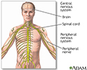
Central nervous system - illustration
The central nervous system is comprised of the brain and spinal cord. The peripheral nervous system includes all peripheral nerves.
Central nervous system
illustration
Review Date: 7/4/2016
Reviewed By: Amit M. Shelat, DO, FACP, Attending Neurologist and Assistant Professor of Clinical Neurology, SUNY Stony Brook, School of Medicine, Stony Brook, NY. Review provided by VeriMed Healthcare Network. Also reviewed by David Zieve, MD, MHA, Isla Ogilvie, PhD, and the A.D.A.M. Editorial team.

