Cherry angioma
Angioma - cherry; Senile angioma; Campbell de Morgan spots; de Morgan spots
A cherry angioma is a noncancerous (benign) skin growth made up of blood vessels.
Causes
Cherry angiomas are fairly common skin growths that vary in size. They can occur almost anywhere on the body, but usually develop on the trunk.
They are most common after age 30. The cause is unknown, but they tend to be inherited (genetic).
Symptoms
A cherry angioma is:
- Bright cherry-red
- Small -- pinhead size to about one quarter inch (0.5 centimeter) in diameter
- Smooth, or can stick out from the skin
Exams and Tests
Your health care provider will look at the growth on your skin to diagnose a cherry angioma. No further tests are usually necessary. Sometimes a skin biopsy may be used to confirm the diagnosis.
Skin biopsy
A skin lesion biopsy is when a small amount of skin is removed so it can be examined. The skin is tested to look for skin conditions or diseases. A...

Treatment
Cherry angiomas usually do not need to be treated. If they affect your appearance or bleed often, they may be removed by:
- Burning (electrosurgery/cautery)
-
Freezing (
cryotherapy
)
Cryotherapy
Cryotherapy is a method of superfreezing tissue in order to destroy it. This article discusses cryotherapy of the skin.
Read Article Now Book Mark Article - Laser
-
Shave excision
Shave excision
A skin lesion is an area of the skin that is different than the surrounding skin. This can be a lump, sore, or an area of skin that is not normal. ...
Read Article Now Book Mark Article
Outlook (Prognosis)
Cherry angiomas are noncancerous. They usually do not harm your health. Removal usually does not cause scarring.
Possible Complications
A cherry angioma may cause:
- Bleeding if it is injured
- Changes in appearance
- Emotional distress
When to Contact a Medical Professional
Call your provider if:
- You have symptoms of a cherry angioma and you would like to have it removed
- The appearance of a cherry angioma (or any skin lesion) changes
References
Habif TP. Vascular tumors and malformations. In: Habif TP, ed. Clinical Dermatology . 6th ed. Philadelphia, PA: Elsevier; 2016:chap 23.
Patterson JW. Vascular tumors. In: Patterson JW, ed. Weedon's Skin Pathology . 4th ed. Philadelphia, PA: Elsevier; 2016:chap 38.
-
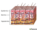
Skin layers - illustration
The skin is the largest organ of the body. The skin and its derivatives (hair, nails, sweat and oil glands) make up the integumentary system. One of the main functions of the skin is protection. It protects the body from external factors such as bacteria, chemicals, and temperature. The skin contains secretions that can kill bacteria and the pigment melanin provides a chemical pigment defense against ultraviolet light that can damage skin cells. Another important function of the skin is body temperature regulation. When the skin is exposed to a cold temperature, the blood vessels in the dermis constrict. This allows the blood which is warm, to bypass the skin. The skin then becomes the temperature of the cold it is exposed to. Body heat is conserved since the blood vessels are not diverting heat to the skin anymore. Among its many functions the skin is an incredible organ always protecting the body from external agents.
Skin layers
illustration
-
Skin layers - illustration
The skin is the largest organ of the body. The skin and its derivatives (hair, nails, sweat and oil glands) make up the integumentary system. One of the main functions of the skin is protection. It protects the body from external factors such as bacteria, chemicals, and temperature. The skin contains secretions that can kill bacteria and the pigment melanin provides a chemical pigment defense against ultraviolet light that can damage skin cells. Another important function of the skin is body temperature regulation. When the skin is exposed to a cold temperature, the blood vessels in the dermis constrict. This allows the blood which is warm, to bypass the skin. The skin then becomes the temperature of the cold it is exposed to. Body heat is conserved since the blood vessels are not diverting heat to the skin anymore. Among its many functions the skin is an incredible organ always protecting the body from external agents.
Skin layers
illustration
Review Date: 10/24/2016
Reviewed By: David L. Swanson, MD, Vice Chair of Medical Dermatology, Associate Professor of Dermatology, Mayo Medical School, Scottsdale, AZ. Also reviewed by David Zieve, MD, MHA, Isla Ogilvie, PhD, and the A.D.A.M. Editorial team.