MRI
Magnetic resonance imaging; Nuclear magnetic resonance (NMR) imaging
A magnetic resonance imaging (MRI) scan is an imaging test that uses powerful magnets and radio waves to create pictures of the body. It does not use radiation (x-rays).
Single MRI images are called slices. The images can be stored on a computer or printed on film. One exam produces dozens or sometimes hundreds of images.
Different types of MRI include:
-
Abdominal MRI
Abdominal MRI
An abdominal magnetic resonance imaging scan is an imaging test that uses powerful magnets and radio waves. The waves create pictures of the inside ...
 ImageRead Article Now Book Mark Article
 -
Cervical MRI
Cervical MRI
A cervical MRI (magnetic resonance imaging) scan uses energy from strong magnets to create pictures of the part of the spine that runs through the ne...
Read Article Now Book Mark Article -
Chest MRI
Chest MRI
A chest MRI (magnetic resonance imaging) scan is an imaging test that uses powerful magnetic fields and radio waves to create pictures of the chest (...
 ImageRead Article Now Book Mark Article
 -
Cranial MRI
Cranial MRI
A head MRI (magnetic resonance imaging) is an imaging test that uses powerful magnets and radio waves to create pictures of the brain and surrounding...
 ImageRead Article Now Book Mark Article
 -
Heart MRI
Heart MRI
Heart magnetic resonance imaging is an imaging method that uses powerful magnets and radio waves to create pictures of the heart. It does not use ra...
 ImageRead Article Now Book Mark Article
 -
Lumbar MRI
Lumbar MRI
A lumbar magnetic resonance imaging (MRI) scan uses energy from strong magnets to create pictures of the lower part of the spine (lumbar spine). An M...
Read Article Now Book Mark Article -
Pelvic MRI
Pelvic MRI
A pelvis MRI (magnetic resonance imaging) scan is a imaging test that uses a machine with powerful magnets and radio waves to create pictures of the ...
Read Article Now Book Mark Article -
MRA (MR Angiography)
MRA (MR Angiography)
Magnetic resonance angiography (MRA) is an MRI exam of the blood vessels. Unlike traditional angiography that involves placing a tube (catheter) int...
Read Article Now Book Mark Article - MRV (MR Venography)
How the Test is Performed
You may be asked to wear a hospital gown or clothing without zippers or snaps (such as sweatpants and a t-shirt). Certain types of metal can cause blurry images.
You will lie on a narrow table, which slides into a large tunnel-shaped scanner.
Some exams require a special dye (contrast). Most of the time, the dye will be given through a vein (IV) in your hand or forearm before the test. The dye helps the radiologist see certain areas more clearly.
Small devices, called coils, may be placed around the head, arm, or leg, or around other areas to be studied. These help send and receive the radio waves, and improve the quality of the images.
During the MRI, the person who operates the machine will watch you from another room. The test lasts about 30 to 60 minutes, but may take longer.
How to Prepare for the Test
You may be asked not to eat or drink anything for 4 to 6 hours before the scan.
Tell your health care provider if you are afraid of close spaces (have claustrophobia). You may be given a medicine to help you feel sleepy and less anxious, or your provider may suggest an open MRI, in which the machine is not as close to the body.
Before the test, tell your provider if you have:
- Artificial heart valves
- Brain aneurysm clips
- Heart defibrillator or pacemaker
- Inner ear (cochlear) implants
- Kidney disease or dialysis (you may not be able to receive contrast)
- Recently placed artificial joints
-
Vascular
stents
Stents
A stent is a tiny tube placed into a hollow structure in your body. This structure can be an artery, a blood vessel, or something such as the tube t...
 ImageRead Article Now Book Mark Article
 - Worked with sheet metal in the past (you may need tests to check for metal pieces in your eyes)
Because the MRI contains strong magnets, metal objects are not allowed into the room with the MRI scanner:
- Items such as jewelry, watches, credit cards, and hearing aids can be damaged.
- Pens, pocketknives, and eyeglasses may fly across the room.
- Pins, hairpins, metal zippers, and similar metallic items can distort the images.
- Removable dental work should be taken out just before the scan.
How the Test will Feel
An MRI exam causes no pain. If you have difficulty lying still or are very nervous, you may be given a medicine to relax you. Too much movement can blur MRI images and cause errors.
The table may be hard or cold, but you can request a blanket or pillow. The machine produces loud thumping and humming noises when turned on. You can wear ear plugs to help reduce the noise.
An intercom in the room allows you to speak to someone at any time. Some MRIs have televisions and special headphones that you can use to help the time pass.
There is no recovery time, unless you were given a medicine to relax. After an MRI scan, you can resume your normal diet, activity, and medicines.
Why the Test is Performed
Having MRIs with other imaging methods can often help your provider make a diagnosis.
MRI images taken after a special dye (contrast) is delivered into your body may provide extra information about the blood vessels.
A magnetic resonance angiogram (MRA) , is a form of magnetic resonance imaging that creates 3-dimensional pictures of blood vessels. It is often used when traditional angiography cannot be done.
Magnetic resonance angiogram (MRA)
Magnetic resonance angiography (MRA) is an MRI exam of the blood vessels. Unlike traditional angiography that involves placing a tube (catheter) int...
Angiography
An arteriogram is an imaging test that uses x-rays and a special dye to see inside the arteries. It can be used to view arteries in the heart, brain...

Normal Results
A normal result means the body area being studied looks normal.
What Abnormal Results Mean
Results depend on the part of the body being examined and the nature of the problem. Different types of tissues send back different MRI signals. For example, healthy tissue sends back a slightly different signal than cancerous tissue. Consult your provider with any questions and concerns.
Risks
MRI does not use ionizing radiation. No side effects from the magnetic fields and radio waves have been reported.
The most common type of contrast (dye) used is gadolinium. It is very safe. Allergic reactions rarely occur. However, gadolinium can be harmful to people with kidney problems who are on dialysis. Tell your provider before the test if you have kidney problems.
The strong magnetic fields created during an MRI can cause heart pacemakers and other implants not to work as well. The magnets can also cause a piece of metal inside your body to move or shift.
References
Litt H, Carpenter JP. Magnetic resonance imaging. In: Cronenwett JL, Johnston KW, eds. Rutherford's Vascular Surgery . 8th ed. Philadelphia, PA: Elsevier Saunders; 2014:chap 23.
Wahl RL. Imaging. In: Niederhuber JE, Armitage JO, Doroshow JH, Kastan MB, Tepper JE, eds. Abeloff's Clinical Oncology . 5th ed. Philadelphia, PA: Elsevier Churchill Livingstone; 2014:chap 18.
Wilkinson ID, Graves MJ. Magnetic resonance imaging. In: Adam A, Dixon AK, Gillard JH, Schaefer-Prokop CM, eds. Grainger & Allison's Diagnostic Radiology. 6th ed. New York, NY: Elsevier; 2015:chap 5.
-
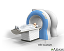
MRI scans - illustration
MRI stands for magnetic resonance imaging. It allows imaging of the interior of the body without using x-rays or other types of ionizing radiation. An MRI scan is capable of showing fine detail of different tissues.
MRI scans
illustration
Review Date: 7/3/2016
Reviewed By: Jason Levy, MD, Northside Radiology Associates, Atlanta, GA. Also reviewed by David Zieve, MD, MHA, Isla Ogilvie, PhD, and the A.D.A.M. Editorial team.