Intravascular ultrasound
IVUS; Ultrasound - coronary artery; Endovascular ultrasound; Intravascular echocardiography
Intravascular ultrasound (IVUS) is a diagnostic test. This test uses sound waves to see inside blood vessels. It is useful for evaluating the coronary arteries that supply the heart.
Description
A tiny ultrasound wand is attached to the top of a thin tube. This tube is called a catheter. The catheter is inserted into an artery in your groin area and moved up to the heart. It is different from conventional Duplex ultrasound. Duplex ultrasound is done from the outside of your body by placing the transducer on the skin.
Ultrasound
Ultrasound uses high-frequency sound waves to make images of organs and structures inside the body.

A computer measures how the sound waves reflect off blood vessels, and changes the sound waves into pictures. IVUS gives the health care provider a look at your coronary arteries from the inside-out.
IVUS is almost always done during a procedure. Reasons why it may be done include:
-
Getting information about the heart or its blood vessels
or to find out if you need heart surgery
Getting information about the heart or ...
Cardiac catheterization involves passing a thin flexible tube (catheter) into the right or left side of the heart. The catheter is most often insert...
 ImageRead Article Now Book Mark Article
 -
Treating some types of heart conditions
Treating some types of heart conditions
Angioplasty is a procedure to open narrowed or blocked blood vessels that supply blood to the heart. These blood vessels are called the coronary art...
 ImageRead Article Now Book Mark Article
Angiography gives a general look at the coronary arteries. However, it can't show the walls of the arteries. IVUS images show the artery walls and can reveal cholesterol and fat deposits (plaques). Buildup of these deposits can increase your risk of a heart attack.
IVUS has helped providers understand how stents become clogged. This is called stent restenosis.
Stents
A stent is a tiny tube placed into a hollow structure in your body. This structure can be an artery, a blood vessel, or something such as the tube t...
Why the Procedure Is Performed
IVUS is commonly done to make sure a stent is correctly placed during angioplasty. It may also be done to determine where a stent should be placed.
IVUS may also be used to:
- View the aorta and structure of the artery walls, which can show plaque buildup
- Find which blood vessel is involved in aortic dissection
Risks
There is a slight risk of complications with angioplasty and cardiac catheterization. However, the tests are very safe when done by an experienced team. IVUS adds little additional risk.
Risks of anesthesia and surgery in general are:
- Reactions to medicines
- Breathing problems
- Bleeding, blood clots
- Infection
Other risks include:
- Damage to a heart valve or blood vessel
-
Heart attack
Heart attack
Most heart attacks are caused by a blood clot that blocks one of the coronary arteries. The coronary arteries bring blood and oxygen to the heart. ...
 ImageRead Article Now Book Mark Article
 -
Irregular heartbeat (
arrhythmia
)
Arrhythmia
An arrhythmia is a disorder of the heart rate (pulse) or heart rhythm. The heart can beat too fast (tachycardia), too slow (bradycardia), or irregul...
ImageRead Article Now Book Mark Article - Kidney failure (a higher risk in people who already have kidney problems or diabetes)
-
Stroke
(this is rare)
Stroke
A stroke occurs when blood flow to a part of the brain stops. A stroke is sometimes called a "brain attack. " If blood flow is cut off for longer th...
 ImageRead Article Now Book Mark Article
After the Procedure
After the test, the catheter is completely removed. A bandage is placed on the area. You will be asked to lie flat on your back with pressure on your groin area for a few hours after the test to prevent bleeding.
If IVUS was done during:
- Cardiac catheterization: You will stay in the hospital for about 3 to 6 hours.
- Angioplasty: You will stay in the hospital for 12 to 24 hours.
The IVUS does not add to the time you must stay in the hospital.
References
American Heart Association. Cardiac catheterization. Updated September 2, 2015. www.heart.org/HEARTORG/Conditions/HeartAttack/SymptomsDiagnosisofHeartAttack/Cardiac-Catheterization_UCM_451486_Article.jsp#.V34vl7h961s . Accessed July 7, 2016.
León LR, Labropoulos N. Vascular laboratory: venous duplex scanning. In: Cronenwett JL, Johnston KW, eds. Rutherford's Vascular Surgery. 8th ed. Philadelphia, PA: Elsevier Saunders; 2014:chap 18.
Ziada KM. Intravascular ultrasound imaging. In: Bhatt DL, ed. Cardiovascular Intervention: A Companion to Braunwald's Heart Disease . Philadelphia, PA: Elsevier; 2016:chap 16.
-
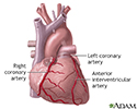
Anterior heart arteries - illustration
The coronary arteries supply blood to the heart muscle. The right coronary artery supplies both the left and the right heart; the left coronary artery supplies the left heart.
Anterior heart arteries
illustration
-
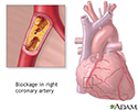
Coronary artery blockage - illustration
Atherosclerosis is a common disorder of the arteries. Fat, cholesterol, and other substances collect in the walls of arteries. Larger accumulations are called atheromas or plaque and can damage artery walls and block blood flow. Severely restricted blood flow in the heart muscle leads to symptoms such as chest pain.
Coronary artery blockage
illustration
-
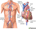
Cardiac catheterization - illustration
Cardiac catheterization is used to study the various functions of the heart. Using different techniques, the coronary arteries can be viewed by injecting dye or opened using balloon angioplasty. The oxygen concentration can be measured across the valves and walls (septa) of the heart and pressures within each chamber of the heart and across the valves can be measured. The technique can even be performed in small, newborn infants.
Cardiac catheterization
illustration
-
Anterior heart arteries - illustration
The coronary arteries supply blood to the heart muscle. The right coronary artery supplies both the left and the right heart; the left coronary artery supplies the left heart.
Anterior heart arteries
illustration
-
Coronary artery blockage - illustration
Atherosclerosis is a common disorder of the arteries. Fat, cholesterol, and other substances collect in the walls of arteries. Larger accumulations are called atheromas or plaque and can damage artery walls and block blood flow. Severely restricted blood flow in the heart muscle leads to symptoms such as chest pain.
Coronary artery blockage
illustration
-
Cardiac catheterization - illustration
Cardiac catheterization is used to study the various functions of the heart. Using different techniques, the coronary arteries can be viewed by injecting dye or opened using balloon angioplasty. The oxygen concentration can be measured across the valves and walls (septa) of the heart and pressures within each chamber of the heart and across the valves can be measured. The technique can even be performed in small, newborn infants.
Cardiac catheterization
illustration
Review Date: 6/6/2016
Reviewed By: Deepak Sudheendra, MD, RPVI, Assistant Professor of Interventional Radiology and Surgery at the University of Pennsylvania Perelman School of Medicine, with an expertise in Vascular Interventional Radiology and Surgical Critical Care, Philadelphia, PA. Review provided by VeriMed Healthcare Network. Also reviewed by David Zieve, MD, MHA, Isla Ogilvie, PhD, and the A.D.A.M. Editorial team.