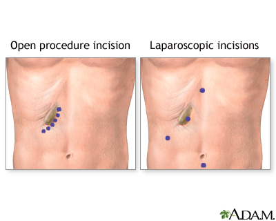
Gallbladder removal - open
Cholecystectomy - open; Surgery - gallbladder - open
Open gallbladder removal is surgery to remove the gallbladder through a large cut in your abdomen.
Description
Surgery is done while you are under general anesthesia so you will be asleep and pain-free. To perform the surgery:
General anesthesia
General anesthesia is treatment with certain medicines that puts you into a deep sleep so you do not feel pain during surgery. After you receive the...
- The surgeon makes a 5 to 7 inch (12.5 to 17.5 centimeters) cut in the upper right part of your belly, just below your ribs.
- The area is opened up so the surgeon can view the gallbladder and separate it from the other organs.
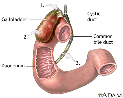
- The surgeon cuts the bile duct and blood vessels that lead to the gallbladder.
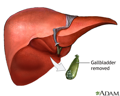
- The gallbladder is gently lifted out and removed from your body.
An x-ray called a cholangiogram may be done during your surgery.
Cholangiogram
A percutaneous transhepatic cholangiogram (PTCA) is an x-ray of the bile ducts. These are the tubes that carry bile from the liver to the gallbladde...

- To do this test, dye is injected into your common bile duct and an x-ray is taken. The dye helps find stones that may be outside your gallbladder.
- If other stones are found, the surgeon may remove them with a special instrument.
The surgery takes about 1 hour.
Why the Procedure Is Performed
You may need this surgery if you have pain or other symptoms from gallstones. You may also need surgery if your gallbladder is not working normally.
Common symptoms may include:
-
Indigestion
, including bloating, heartburn, and gas
Indigestion
Indigestion (dyspepsia) is a mild discomfort in the upper belly or abdomen. It occurs during or right after eating. It may feel like:Heat, burning,...
 ImageRead Article Now Book Mark Article
 -
Nausea and vomiting
Nausea and vomiting
Nausea is feeling an urge to vomit. It is often called "being sick to your stomach. "Vomiting or throwing-up is forcing the contents of the stomach ...
 ImageRead Article Now Book Mark Article
 -
Pain after eating, usually in the upper right or upper middle area of your
belly
(epigastric pain)
Belly
Abdominal pain is pain that you feel anywhere between your chest and groin. This is often referred to as the stomach region or belly.
 ImageRead Article Now Book Mark Article
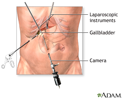
The most common way to remove the gallbladder is by using a medical instrument called a laparoscope ( laparoscopic cholecystectomy ). Open gallbladder surgery is used when laparoscopic surgery cannot be done safely. In some cases, the surgeon needs to switch to an open surgery if laparoscopic surgery cannot be successfully continued.
Laparoscopic cholecystectomy
Laparoscopic gallbladder removal is surgery to remove the gallbladder using a medical device called a laparoscope.

Other reasons for removing the gallbladder by open surgery:
- Unexpected bleeding during the laparoscopic operation
-
Obesity
Obesity
Obesity means having too much body fat. It is not the same as being overweight, which means weighing too much. A person may be overweight from extr...
 ImageRead Article Now Book Mark Article
 - Pancreatitis (inflammation in the pancreas)
- Pregnancy (third trimester)
-
Severe
liver problems
Liver problems
The term "liver disease" applies to many conditions that stop the liver from working or prevent it from functioning well. Abdominal pain, yellowing ...
 ImageRead Article Now Book Mark Article
 - Past surgeries in the same area of your belly
Risks
Risks of anesthesia and surgery in general are:
- Reactions to medicines
-
Breathing problems
Breathing problems
Breathing difficulty may involve:Difficult breathingUncomfortable breathingFeeling like you are not getting enough air
 ImageRead Article Now Book Mark Article
 -
Bleeding,
blood clots
Blood clots
Blood clots are clumps that occur when blood hardens from a liquid to a solid. A blood clot that forms inside one of your veins or arteries is calle...
 ImageRead Article Now Book Mark Article
 - Infection
Risks of gallbladder surgery are:
- Damage to the blood vessels that go to the liver
- Injury to the common bile duct
- Injury to the small or large intestine
- Pancreatitis (inflammation of the pancreas)
Before the Procedure
Your may have the following tests done before surgery:
-
Blood tests (
complete blood count
,
electrolytes
, liver and kidney tests)
Complete blood count
A complete blood count (CBC) test measures the following:The number of red blood cells (RBC count)The number of white blood cells (WBC count)The tota...
 ImageRead Article Now Book Mark Article
Electrolytes
The electrolytes - urine test measures specific chemicals called electrolytes in urine. It usually measures the levels of calcium, chloride, potassi...
 ImageRead Article Now Book Mark Article
 -
Chest x-ray
or electrocardiogram (
EKG
), for some patients
Chest x-ray
A chest x-ray is an x-ray of the chest, lungs, heart, large arteries, ribs, and diaphragm.
 ImageRead Article Now Book Mark Article
EKG
An electrocardiogram (ECG) is a test that records the electrical activity of the heart.
 ImageRead Article Now Book Mark Article
 - Several x-rays of the gallbladder
-
Ultrasound
of the gallbladder
Ultrasound
Ultrasound uses high-frequency sound waves to make images of organs and structures inside the body.
 ImageRead Article Now Book Mark Article
Tell your doctor or nurse:
- If you are or might be pregnant
- Which drugs, vitamins, and other supplements you are taking, even ones you bought without a prescription
During the week before surgery:
- You may be asked to stop taking aspirin, ibuprofen (Advil, Motrin), vitamin E, warfarin (Coumadin), and any other drugs that may put you at a higher risk of bleeding during surgery.
- Ask your doctor which drugs you should still take on the day of your surgery.
-
Prepare your home
for any problems you might have getting around after the surgery.
Prepare your home
Getting your home ready after you have been in the hospital often requires much preparation. Set up your home to make your life is easier and safer w...
Read Article Now Book Mark Article - You'll be told when to arrive at the hospital.
On the day of surgery:
- Follow instructions about when to stop eating and drinking.
- Take the drugs your doctor told you to take with a small sip of water.
- Shower the night before or the morning of your surgery.
- Arrive at the hospital on time.
After the Procedure
You may stay in the hospital for 3 to 5 days after open gallbladder removal. During that time:
- You may be asked to breathe into a device called an incentive spirometer. This helps keep your lungs working well so that you do not get pneumonia.
- The nurse will help you sit up in bed, hang your legs over the side, and then stand up and start to walk.
- At first, you will receive fluids into your vein through an intravenous (IV) tube. Soon after, you will be asked to start drinking liquids and eating foods.
- You will be able to shower while you are still in the hospital.
- You may be asked to wear pressure stockings on your legs to help prevent a blood clot from forming. These stockings also help keep your blood circulating well.
If there were problems during your surgery, or if you have bleeding, a lot of pain, or a fever, you may need to stay in the hospital longer. Your doctor or nurses will tell you how to care for yourself after you leave the hospital.
How to care for yourself
Cholelithiasis - open discharge; Biliary calculus - open discharge; Gallstones - open discharge; Cholecystitis - open discharge; Cholecystectomy - op...

Outlook (Prognosis)
Most people recover quickly and have good results from this procedure.
References
Glasgow RE, Mulvihill SJ. Treatment of gallstone disease: In: Feldman M, Friedman LS, Brandt LJ, eds. Sleisenger and Fordtran's Gastrointestinal and Liver Disease Pathophysiology/Diagnosis/Management . 9th ed. Philadelphia, PA: Elsevier Saunders; 2010:chap 66.
Jackson PG, Evans SRT. Biliary system. In: Townsend CM Jr, Beauchamp RD, Evers BM, Mattox KL, eds. Sabiston Textbook of Surgery . 19th ed. Philadelphia, PA: Elsevier Saunders; 2012:chap 55.
-
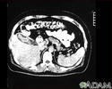
Cholecystitis, CT scan - illustration
This is a CT scan of the upper abdomen showing cholecystitis (gall stones).
Cholecystitis, CT scan
illustration
-
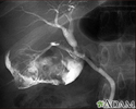
Cholecystitis, cholangiogram - illustration
Cholelithiasis can be seen on a cholangiogram. Radio-opaque dye is used to enhance the x-ray. Multiple stones are present in the gallbladder (PTCA).
Cholecystitis, cholangiogram
illustration
-
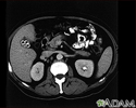
Cholecystolithiasis - illustration
Cholecystolithiasis. CT scan of the upper abdomen showing multiple gallstones.
Cholecystolithiasis
illustration
-
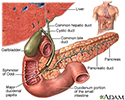
Gallbladder - illustration
The gallbladder is a muscular sac located under the liver. It stores and concentrates the bile produced in the liver that is not immediately needed for digestion. Bile is released from the gallbladder into the small intestine in response to food. The pancreatic duct joins the common bile duct at the small intestine adding enzymes to aid in digestion.
Gallbladder
illustration
-
Gallbladder removal - series
Presentation
-
Gallstones and gallbladder disease
(In-Depth)
Review Date: 7/28/2015
Reviewed By: Debra G. Wechter, MD, FACS, general surgery practice specializing in breast cancer, Virginia Mason Medical Center, Seattle, WA. Also reviewed by David Zieve, MD, MHA, Isla Ogilvie, PhD, and the A.D.A.M. Editorial team.
