CBC blood test
Complete blood count
A complete blood count (CBC) test measures the following:
-
The number of red blood cells (
RBC count
)
RBC count
An RBC count is a blood test that measures how many red blood cells (RBCs) you have. RBCs contain hemoglobin, which carries oxygen. How much oxygen ...
 ImageRead Article Now Book Mark Article
 -
The number of white blood cells (
WBC count
)
WBC count
A WBC count is a blood test to measure the number of white blood cells (WBCs) in the blood. WBCs help fight infections. They are also called leukocy...
 ImageRead Article Now Book Mark Article
 -
The total amount of
hemoglobin
in the blood
Hemoglobin
Hemoglobin is a protein in red blood cells that carries oxygen. The hemoglobin test measures how much hemoglobin is in your blood.
 ImageRead Article Now Book Mark Article
 -
The fraction of the blood composed of red blood cells (
hematocrit
)
Hematocrit
Hematocrit is a blood test that measures how much of a person's blood is made up of red blood cells. This measurement depends on the number of and s...
 ImageRead Article Now Book Mark Article
The CBC test also provides information about the following measurements:
- Average red blood cell size (MCV)
-
Hemoglobin
amount per red blood cell (MCH)
Hemoglobin
Hemoglobin is a protein in red blood cells that carries oxygen. The hemoglobin test measures how much hemoglobin is in your blood.
ImageRead Article Now Book Mark Article - The amount of hemoglobin relative to the size of the cell (hemoglobin concentration) per red blood cell (MCHC)
The platelet count is also usually included in the CBC.
Platelet count
A platelet count is a lab test to measure how many platelets you have in your blood. Platelets are parts of the blood that help the blood clot. The...
How the Test is Performed
A blood sample is needed.
Blood sample
Venipuncture is the collection of blood from a vein. It is most often done for laboratory testing.
How to Prepare for the Test
There is no special preparation needed.
How the Test will Feel
When the needle is inserted to draw blood, you may feel moderate pain. Some people feel only a prick or stinging. Afterward there may be some throbbing or slight bruising. This soon goes away.
Why the Test is Performed
A complete blood count (CBC) is a commonly performed lab test. It can be used to detect or monitor many different health conditions. Your health care provider may order this test:
- As part of a routine check-up
- If you are having symptoms, such as fatigue, weight loss, fever or other signs of an infection, weakness, bruising, bleeding, or any signs of cancer
- When you are receiving treatments (medicines or radiation) that may change your blood count results
- To monitor a chronic health problem that may change your blood count results, such as chronic kidney disease
Normal Results
Blood counts may vary with altitude. In general, normal results are:
RBC count:
- Male: 4.7 to 6.1 million cells/mcL (4.7 to 6.1 x 10^12/L)
- Female: 4.2 to 5.4 million cells/mcL ( 4.2 to 5.4 x 10^12/L)
WBC count:
- 4,500 to 10,000 cells/mcL (4.5 to 11.0 x10^9/L)
Hematocrit:
- Male: 40.7% to 50.3% (0. 41 to 0.50)
- Female: 36.1% to 44.3% (0.36 to 0.44)
Hemoglobin:
- Male: 13.8 to 17.2 gm/dL (138 to 172 g/L)
- Female: 12.1 to 15.1 gm/dL (121 to 151 g/L)
Red blood cell indices:
- MCV: 80 to 95 femtoliter
- MCH: 27 to 31 pg/cell
- MCHC: 32 to 36 gm/dL (320 to 360 g/L)
Platelet count:
- 150,000 to 450,000/dL (150 to 450 x 10^9/L)
The examples above are common measurements for results of these tests. Normal value ranges may vary slightly among different laboratories. Some labs use different measurements or test different samples. Talk to your doctor about the meaning of your specific test results.
What Abnormal Results Mean
High RBC, hemoglobin, or hematocrit may be due to:
- A lack of enough water and fluids, such as from severe diarrhea, excessive sweating, or water pills used to treat high blood pressure
-
Kidney disease with high
erythropoietin
production
Erythropoietin
The erythropoietin test measures the amount of a hormone called erythropoietin (EPO) in blood. The hormone tells stem cells in the bone marrow to mak...
Read Article Now Book Mark Article - Low oxygen level in the blood for a long time, most often due to heart or lung disease
-
Polycythemia vera
Polycythemia vera
Polycythemia vera is a bone marrow disease that leads to an abnormal increase in the number of blood cells. The red blood cells are mostly affected....
Read Article Now Book Mark Article - Smoking
Low RBC, hemoglobin, or hematacrit is a sign of anemia, which can result from:
-
Blood loss
(either sudden, or from problems such as heavy menstrual periods over a long time)
Blood loss
Bleeding is the loss of blood. Bleeding may be:Inside the body (internally) Outside the body (externally)Bleeding may occur:Inside the body when blo...
 ImageRead Article Now Book Mark Article
 - Bone marrow failure (for example, from radiation, infection, or tumor)
- Breakdown of red blood cells ( hemolysis )
- Cancer and cancer treatment
- Certain long-term (chronic) medical conditions, such as chronic kidney disease, ulcerative colitis, or rheumatoid arthritis
- Leukemia
-
Long-term infections such as
hepatitis
Hepatitis
Hepatitis is swelling and inflammation of the liver.
 ImageRead Article Now Book Mark Article
 -
Poor diet and nutrition, causing too little iron,
folate
,
vitamin B12
, or
vitamin B6
Folate
Folic acid is a type of B vitamin. It is the man-made (synthetic) form of folate that is found in supplements and added to fortified foods. Folate i...
 ImageRead Article Now Book Mark Article
Vitamin B12
Vitamin B12 is a water-soluble vitamin. Water-soluble vitamins dissolve in water. After the body uses these vitamins, leftover amounts leave the bo...
 ImageRead Article Now Book Mark Article
Vitamin B6
Vitamin B6 is a water-soluble vitamin. Water-soluble vitamins dissolve in water so the body cannot store them. Leftover amounts of the vitamin leav...
 ImageRead Article Now Book Mark Article
 -
Multiple myeloma
Multiple myeloma
Multiple myeloma is a blood cancer that starts in the plasma cells in the bone marrow. Bone marrow is the soft, spongy tissue found inside most bone...
 ImageRead Article Now Book Mark Article
A lower than normal white blood cell count is called leukopenia. A decreased WBC count may be due to:
- Alcohol abuse and liver damage
-
Autoimmune diseases (such as
systemic lupus erythematosus
)
Systemic lupus erythematosus
Systemic lupus erythematosus (SLE) is an autoimmune disease. In this disease, the body's immune system mistakenly attacks healthy tissue. It can af...
 ImageRead Article Now Book Mark Article
 - Bone marrow failure (for example, due to infection, tumor, radiation, or fibrosis)
- Chemotherapy medicines used to treat cancer
- Disease of the liver or spleen
- Enlarged spleen
- Infections caused by viruses, such as mono or AIDS
- Medications
A high WBC count is called leukocytosis. It can result from:
- Certain medicines, such as corticosteroids
- Infections
-
Diseases such as lupus,
rheumatoid arthritis
or
allergy
Rheumatoid arthritis
Rheumatoid arthritis (RA) is a long-term disease. It leads to inflammation of the joints and surrounding tissues. It can also affect other organs....
 ImageRead Article Now Book Mark Article
Allergy
An allergy is an immune response or reaction to substances that are usually not harmful.
 ImageRead Article Now Book Mark Article
 - Leukemia
- Severe emotional or physical stress
- Tissue damage (such as from burns or a heart attack)
A high platelet count may be due to:
- Bleeding
- Diseases such as cancer
- Iron deficiency
- Problems with the bone marrow
A low platelet count may be due to:
- Anemia (various types)
- Disorders where platelets are destroyed
- Pregnancy
- Enlarged spleen
- Bone marrow failure (for example, due to infection, tumor, radiation, or fibrosis)
- Chemotherapy medicines used to treat cancer
Risks
There is very little risk involved with having your blood taken. Veins and arteries vary in size from one person to another and from one side of the body to the other. Taking blood from some people may be more difficult than from others.
Other risks associated with having blood drawn are slight but may include:
- Excessive bleeding
- Fainting or feeling light-headed
- Hematoma (blood accumulating under the skin)
- Infection (a slight risk any time the skin is broken)
Considerations
RBCs transport hemoglobin which, in turn, carries oxygen. The amount of oxygen received by body tissues depends on the amount and function of RBCs and hemoglobin.
WBCs are mediators of inflammation and the immune response . There are various types of WBCs that normally appear in the blood:
Immune response
The immune response is how your body recognizes and defends itself against bacteria, viruses, and substances that appear foreign and harmful....

- Neutrophils (polymorphonuclear leukocytes)
- Band cells (slightly immature neutrophils)
- T-type lymphocytes (T cells)
- B-type lymphocytes (B cells)
- Monocytes
-
Eosinophils
Eosinophils
An absolute eosinophil count is a blood test that measures the number of white blood cells called eosinophils. Eosinophils become active when you ha...
 ImageRead Article Now Book Mark Article
 - Basophils
References
Vajpayee N, Graham SS, Bem S. Basic examination of blood and bone marrow. In: McPherson RA, Pincus MR, eds. Henry's Clinical Diagnosis and Management by Laboratory Methods . 22nd ed. Philadelphia, PA: Elsevier Saunders; 2011:chap 30.
-
Red blood cells, sickle cell - illustration
Sickle cell anemia is an inherited blood disease in which the red blood cells produce abnormal pigment (hemoglobin). The abnormal hemoglobin causes deformity of the red blood cells into crescent or sickle-shapes, as seen in this photomicrograph.
Red blood cells, sickle cell
illustration
-
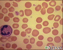
Megaloblastic anemia - view of red blood cells - illustration
This picture shows large, dense, oversized, red blood cells (RBCs) that are seen in megaloblastic anemia. Megaloblastic anemia can occur when there is a deficiency of vitamin B-12.
Megaloblastic anemia - view of red blood cells
illustration
-
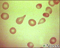
Red blood cells, tear-drop shape - illustration
This photomicrograph shows one of the abnormal shapes that red blood cells (RBCs) may assume, a tear-drop shape. Normally, RBCs are round.
Red blood cells, tear-drop shape
illustration
-
Red blood cells, normal - illustration
This photomicrograph shows normal red blood cells (RBCs) as seen in the microscope after staining.
Red blood cells, normal
illustration
-
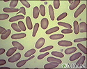
Red blood cells, elliptocytosis - illustration
Elliptocytosis is a hereditary disorder of the red blood cells (RBCs). In this condition, the RBCs assume an elliptical shape, rather than the typical round shape.
Red blood cells, elliptocytosis
illustration
-
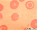
Red blood cells, spherocytosis - illustration
Spherocytosis is a hereditary disorder of the red blood cells (RBCs), which may be associated with a mild anemia. Typically, the affected RBCs are small, spherically shaped, and lack the light centers seen in normal, round RBCs.
Red blood cells, spherocytosis
illustration
-
Red blood cells, multiple sickle cells - illustration
Sickle cell anemia is an inherited disorder in which abnormal hemoglobin (the red pigment inside red blood cells) is produced. The abnormal hemoglobin causes red blood cells to assume a sickle shape, like the ones seen in this photomicrograph.
Red blood cells, multiple sickle cells
illustration
-
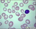
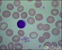
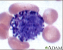
Basophil (close-up) - illustration
Basophils are a specific type of white blood cell. These cells are readily stained with basic dyes (this is where the name comes from). Note the dark grains inside the cellular fluid (cytoplasm) of this basophil. Basophils make up only a small portion of the number of white blood cells but are important parts of the body's immune response. They release histamine and other chemicals that act on the blood vessels when the immune response is triggered.
Basophil (close-up)
illustration
-
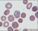
Malaria, microscopic view of cellular parasites - illustration
Malarial parasites are visible within the red blood cells. They are stained a dark bluish color.
Malaria, microscopic view of cellular parasites
illustration
-
Malaria, photomicrograph of cellular parasites - illustration
Malaria is a disease caused by parasites. This picture shows dark orange-stained malaria parasites inside red blood cells (a) and outside the cells (b). Note the large cells that look like targets; it is unknown how these target cells are related to this disease.
Malaria, photomicrograph of cellular parasites
illustration
-
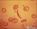
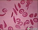
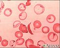
Red blood cells, sickle cells - illustration
These crescent or sickle-shaped red blood cells (RBCs) are present with Sickle cell anemia, and stand out clearly against the normal round RBCs. These abnormally shaped cells may become entangled and block blood flow in the small blood vessels (capillaries).
Red blood cells, sickle cells
illustration
-
Red blood cells, sickle and pappenheimer - illustration
This photomicrograph of red blood cells (RBCs) shows both sickle-shaped and Pappenheimer bodies.
Red blood cells, sickle and pappenheimer
illustration
-
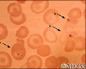
Red blood cells, target cells - illustration
These abnormal red blood cells (RBCs) resemble targets. These cells are seen in association with some forms of anemia, and following the removal of the spleen (splenectomy).
Red blood cells, target cells
illustration
-
Formed elements of blood - illustration
Blood transports oxygen and nutrients to body tissues and returns waste and carbon dioxide. Blood distributes nearly everything that is carried from one area in the body to another place within the body. For example, blood transports hormones from endocrine organs to their target organs and tissues. Blood helps maintain body temperature and normal pH levels in body tissues. The protective functions of blood include clot formation and the prevention of infection.
Formed elements of blood
illustration
-
Complete blood count - series
Presentation

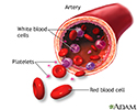
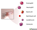
 is a screening test, used to diagnose and manage numerous diseases. It can reflect problems with fluid volume (such as dehydration) or loss of blood. It can show abnormalities in the production, life span, and destruction of blood cells. It can reflect acute or chronic infection, allergies, and problems with clotting. The CBC test isolates and counts the 7 types of cells found in the blood: neutrophil, eosinophil, basophil, red blood cell, lymphocyte, monocyte, and platelet.
</p>")
Multiple punctures to locate veinsInfection (a slight risk any time the skin is broken) Oozing of blood from puncture site Fainting or feeling lightheaded Hematoma (blood accumulating under the skin) Multiple punctures to locate veins Infection (a slight risk any time the skin is broken) </p>")
Erythropoietin deficiency (secondary to kidney disease)Hemolysis (RBC destruction)LeukemiaMultiple myelomaOver hydration Blood loss Iron deficiency Deficiences of vitamin B12 or folic acid Bone marrow failure (for example, from radiation, toxin, fibrosis, tumor) Erythropoietin deficiency (secondary to kidney disease) Hemolysis (RBC destruction) Leukemia Multiple myeloma Over hydration Low numbers of white blood cells (leukopenia) may indicate: Bone marrow failure (for example, due to granuloma (granular tumor), tumor, or fibrosis)Presence of cytotoxic substanceCollagen-vascular diseases (such as lupus erythematosus)Disease of the liver or spleenRadiation exposure Bone marrow failure (for example, due to granuloma (granular tumor), tumor, or fibrosis) Presence of cytotoxic substance Collagen-vascular diseases (such as lupus erythematosus) Disease of the liver or spleen Radiation exposure High numbers of white blood cells (leukocytosis) may indicate: Infectious diseasesInflammatory disease (such as rheumatoid arthritis or allergy)LeukemiaSevere emotional or physical stressTissue damage (for example, burns) Infectious diseases Inflammatory disease (such as rheumatoid arthritis or allergy) Leukemia Severe emotional or physical stress Tissue damage (for example, burns) A high hematocrit may indicate: DehydrationBurnsDiarrheaEclampsiaErythrocytosisPolycythemia veraShock Dehydration Burns Diarrhea Eclampsia Erythrocytosis Polycythemia vera Shock </p>")
Emphysema Cigarette smoking Low oxygen tension in the blood Congenital heart disease Cor pulmonale Pulmonary fibrosis Polycythemia vera Dehydration (such as from severe diarrhea) Emphysema </p>")
-
Red blood cells, sickle cell - illustration
Sickle cell anemia is an inherited blood disease in which the red blood cells produce abnormal pigment (hemoglobin). The abnormal hemoglobin causes deformity of the red blood cells into crescent or sickle-shapes, as seen in this photomicrograph.
Red blood cells, sickle cell
illustration
-
Megaloblastic anemia - view of red blood cells - illustration
This picture shows large, dense, oversized, red blood cells (RBCs) that are seen in megaloblastic anemia. Megaloblastic anemia can occur when there is a deficiency of vitamin B-12.
Megaloblastic anemia - view of red blood cells
illustration
-
Red blood cells, tear-drop shape - illustration
This photomicrograph shows one of the abnormal shapes that red blood cells (RBCs) may assume, a tear-drop shape. Normally, RBCs are round.
Red blood cells, tear-drop shape
illustration
-
Red blood cells, normal - illustration
This photomicrograph shows normal red blood cells (RBCs) as seen in the microscope after staining.
Red blood cells, normal
illustration
-
Red blood cells, elliptocytosis - illustration
Elliptocytosis is a hereditary disorder of the red blood cells (RBCs). In this condition, the RBCs assume an elliptical shape, rather than the typical round shape.
Red blood cells, elliptocytosis
illustration
-
Red blood cells, spherocytosis - illustration
Spherocytosis is a hereditary disorder of the red blood cells (RBCs), which may be associated with a mild anemia. Typically, the affected RBCs are small, spherically shaped, and lack the light centers seen in normal, round RBCs.
Red blood cells, spherocytosis
illustration
-
Red blood cells, multiple sickle cells - illustration
Sickle cell anemia is an inherited disorder in which abnormal hemoglobin (the red pigment inside red blood cells) is produced. The abnormal hemoglobin causes red blood cells to assume a sickle shape, like the ones seen in this photomicrograph.
Red blood cells, multiple sickle cells
illustration
-
Basophil (close-up) - illustration
Basophils are a specific type of white blood cell. These cells are readily stained with basic dyes (this is where the name comes from). Note the dark grains inside the cellular fluid (cytoplasm) of this basophil. Basophils make up only a small portion of the number of white blood cells but are important parts of the body's immune response. They release histamine and other chemicals that act on the blood vessels when the immune response is triggered.
Basophil (close-up)
illustration
-
Malaria, microscopic view of cellular parasites - illustration
Malarial parasites are visible within the red blood cells. They are stained a dark bluish color.
Malaria, microscopic view of cellular parasites
illustration
-
Malaria, photomicrograph of cellular parasites - illustration
Malaria is a disease caused by parasites. This picture shows dark orange-stained malaria parasites inside red blood cells (a) and outside the cells (b). Note the large cells that look like targets; it is unknown how these target cells are related to this disease.
Malaria, photomicrograph of cellular parasites
illustration
-
Red blood cells, sickle cells - illustration
These crescent or sickle-shaped red blood cells (RBCs) are present with Sickle cell anemia, and stand out clearly against the normal round RBCs. These abnormally shaped cells may become entangled and block blood flow in the small blood vessels (capillaries).
Red blood cells, sickle cells
illustration
-
Red blood cells, sickle and pappenheimer - illustration
This photomicrograph of red blood cells (RBCs) shows both sickle-shaped and Pappenheimer bodies.
Red blood cells, sickle and pappenheimer
illustration
-
Red blood cells, target cells - illustration
These abnormal red blood cells (RBCs) resemble targets. These cells are seen in association with some forms of anemia, and following the removal of the spleen (splenectomy).
Red blood cells, target cells
illustration
-
Formed elements of blood - illustration
Blood transports oxygen and nutrients to body tissues and returns waste and carbon dioxide. Blood distributes nearly everything that is carried from one area in the body to another place within the body. For example, blood transports hormones from endocrine organs to their target organs and tissues. Blood helps maintain body temperature and normal pH levels in body tissues. The protective functions of blood include clot formation and the prevention of infection.
Formed elements of blood
illustration
-
Complete blood count - series
Presentation
-
Anemia
(In-Depth)
Review Date: 11/26/2014
Reviewed By: Yi-Bin Chen, MD, Leukemia/Bone Marrow Transplant Program, Massachusetts General Hospital, Boston, MA. Also reviewed by David Zieve, MD, MHA, Isla Ogilvie, PhD, and the A.D.A.M. Editorial team.