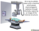
Abdominal x-ray
Abdominal film; X-ray - abdomen; Flat plate; KUB x-ray
An abdominal x-ray is an imaging test to look at organs and structures in the abdomen. Organs include the spleen, stomach, and intestines.
When the test is done to look at the bladder and kidney structures, it is called a KUB (kidneys, ureters, bladder) x-ray.
How the Test is Performed
The test is done in a hospital radiology department. Or it may done in the health care provider's office by an x-ray technologist.
You lie on your back on the x-ray table. The x-ray machine is positioned over your abdominal area. You hold your breath as the picture is taken so that the picture will not be blurry. You may be asked to change position to the side or to stand up for additional pictures.
Men will have a lead shield placed over the testes to protect against the radiation.
How to Prepare for the Test
Before having the x-ray, tell the provider the following:
- If you are pregnant or think you could be pregnant
- Have an IUD inserted
- Have had a barium contrast x-ray in the last 4 days
- If you have taken any medicines such as Pepto Bismol in the last 4 days (this type of medicine can interfere with the x-ray)
You wear a hospital gown during the x-ray procedure. You must remove all jewelry.
How the Test will Feel
There is no discomfort. The x-rays are taken as you lie on your back, side, and while standing.
Why the Test is Performed
Your provider may order this test to:
-
Diagnose a
pain in the abdomen
or unexplained
nausea
Pain in the abdomen
Abdominal pain is pain that you feel anywhere between your chest and groin. This is often referred to as the stomach region or belly.
 ImageRead Article Now Book Mark Article
Nausea
Nausea is feeling an urge to vomit. It is often called "being sick to your stomach. "Vomiting or throwing-up is forcing the contents of the stomach ...
 ImageRead Article Now Book Mark Article
 -
Identify suspected problems in the urinary system, such as a
kidney stone
Kidney stone
A kidney stone is a solid mass made up of tiny crystals. One or more stones can be in the kidney or ureter at the same time.
 ImageRead Article Now Book Mark Article
 - Identify blockage in the intestine
- Locate an object that has been swallowed
- Help diagnose diseases, such as tumors or other conditions
Normal Results
The x-ray will show normal structures for a person your age.
What Abnormal Results Mean
Abnormal findings include:
- Abdominal masses
- Buildup of fluid in the abdomen
-
Certain types of
gallstones
Gallstones
Gallstones are hard deposits that form inside the gallbladder. Gallstones may be as small as a grain of sand or as large as a golf ball.
 ImageRead Article Now Book Mark Article
 - Foreign object in the intestines
- Hole in the stomach or intestines
- Injury to the abdominal tissue
- Intestinal blockage
- Kidney stones
Risks
There is low radiation exposure. X-rays are monitored and regulated to provide the minimum amount of radiation exposure needed to produce the image. Most experts feel that the risk is low compared to the benefits.
Pregnant women and children are more sensitive to the risks of the x-ray. Women should tell their provider if they are, or may be, pregnant.
References
Morrison ID, McLaughlin P, Maher MM. Current status of imaging of the gastrointestinal tract. In: Adam A, Dixon AK, Gillard JH, Schaefer-Prokop CM, eds. Grainger & Allison's Diagnostic Radiology: A Textbook of Medical Imaging. 6th ed. New York, NY: Elsevier Churchill-Livingstone; 2015:chap 25.
-
X-ray - illustration
X-rays are a form of ionizing radiation that can penetrate the body to form an image on film. Structures that are dense (such as bone) will appear white, air will be black, and other structures will be shades of gray depending on density. X-rays can provide information about obstructions, tumors, and other diseases, especially when coupled with the use of barium and air contrast within the bowel.
X-ray
illustration
-
Digestive system - illustration
The esophagus, stomach, large and small intestine, aided by the liver, gallbladder and pancreas convert the nutritive components of food into energy and break down the non-nutritive components into waste to be excreted.
Digestive system
illustration
-
X-ray - illustration
X-rays are a form of ionizing radiation that can penetrate the body to form an image on film. Structures that are dense (such as bone) will appear white, air will be black, and other structures will be shades of gray depending on density. X-rays can provide information about obstructions, tumors, and other diseases, especially when coupled with the use of barium and air contrast within the bowel.
X-ray
illustration
-
Digestive system - illustration
The esophagus, stomach, large and small intestine, aided by the liver, gallbladder and pancreas convert the nutritive components of food into energy and break down the non-nutritive components into waste to be excreted.
Digestive system
illustration
Review Date: 1/31/2015
Reviewed By: Linda J. Vorvick, MD, Medical Director and Director of Didactic Curriculum, MEDEX Northwest Division of Physician Assistant Studies, Department of Family Medicine, UW Medicine, School of Medicine, University of Washington, Seattle, WA. Also reviewed by David Zieve, MD, MHA, Isla Ogilvie, PhD, and the A.D.A.M. Editorial team.


