Orbital cellulitis
Orbital cellulitis is an infection of the fat and muscles around the eye. It affects the eyelids, eyebrows, and cheeks. It may begin suddenly or be a result of an infection that gradually becomes worse.
Causes
Orbital cellulitis is a dangerous infection, which can cause lasting problems. Orbital cellulitis is different than periorbital cellulitis , which is an infection of the eyelid or skin around the eye.
Periorbital cellulitis
Periorbital cellulitis is an infection of the eyelid or skin around the eye.

In children, it often starts out as a bacterial sinus infection from Haemophilus influenza. The infection used to be more common in young children, under the age of 7. It is now rare due to a vaccine that helps prevent this infection.
The bacteria Staphylococcus aureus , Streptococcus pneumoniae , and beta-hemolytic streptococci may also cause orbital cellulitis.
Orbital cellulitis infections in children may get worse very quickly and can lead to blindness. Medical care is needed right away.
Symptoms
Symptoms may include:
- Painful swelling of upper and lower eyelid, and possibly the eyebrow and cheek
-
Bulging eyes
Bulging eyes
Bulging eyes is the abnormal protrusion (bulging out) of one or both eyeballs.
 ImageRead Article Now Book Mark Article
 - Decreased vision
- Pain when moving the eye
- Fever, often 102°F (38.8°C) or higher
- General ill feeling
- Difficult eye movements, perhaps with double vision
- Shiny, red or purple eyelid
Exams and Tests
Tests commonly done include:
-
CBC
(complete blood count)
CBC
A complete blood count (CBC) test measures the following:The number of red blood cells (RBC count)The number of white blood cells (WBC count)The tota...
 ImageRead Article Now Book Mark Article
 -
Blood culture
Blood culture
A blood culture is a laboratory test to check for bacteria or other germs in a blood sample.
Read Article Now Book Mark Article -
Spinal tap
in affected children who are very sick
Spinal tap
Cerebrospinal fluid (CSF) collection is a test to look at the fluid that surrounds the brain and spinal cord. CSF acts as a cushion, protecting the b...
 ImageRead Article Now Book Mark Article
Other tests may include:
-
X-ray of the sinuses
and surrounding area
X-ray of the sinuses
A sinus x-ray is an imaging test to look at the sinuses. These are the air-filled spaces in the front of the skull.
 ImageRead Article Now Book Mark Article
 -
CT scan or MRI of the sinuses and orbit
CT scan or MRI of the sinuses and orbit
A head computed tomography (CT) scan uses many x-rays to create pictures of the head, including the skull, brain, eye sockets, and sinuses.
 ImageRead Article Now Book Mark Article
 - Culture of eye and nose drainage
-
Throat culture
Throat culture
A throat swab culture is a laboratory test that is done to identify germs that may cause infection in the throat. It is most often used to diagnose ...
 ImageRead Article Now Book Mark Article
Treatment
In most cases, a hospital stay is needed. Treatment most often includes antibiotics given through a vein. Surgery may be needed to drain the abscess , or relieve pressure in the space around the eye.
Abscess
An abscess is a collection of pus in any part of the body. In most cases, the area around an abscess is swollen and inflamed.
An orbital cellulitis infection can get worse very quickly. A person with this condition must be checked every few hours.
Outlook (Prognosis)
With prompt treatment, the person can recover fully.
Possible Complications
Complications may include:
-
Cavernous sinus thrombosis
(formation of a blood clot in a cavity at the base of the brain)
Cavernous sinus thrombosis
Cavernous sinus thrombosis is a blood clot in an area at the base of the brain.
ImageRead Article Now Book Mark Article -
Hearing loss
Hearing loss
Hearing loss is being partly or totally unable to hear sound in one or both ears.
 ImageRead Article Now Book Mark Article
 -
Septicemia
or blood infection
Septicemia
Septicemia is bacteria in the blood (bacteremia) that often occurs with severe infections. Also called sepsis, septicemia is a serious, life-threate...
Read Article Now Book Mark Article -
Meningitis
Meningitis
Meningitis is an infection of the membranes covering the brain and spinal cord. This covering is called the meninges.
 ImageRead Article Now Book Mark Article
 -
Optic nerve damage and
loss of vision
Loss of vision
Blindness is a lack of vision. It may also refer to a loss of vision that cannot be corrected with glasses or contact lenses. Partial blindness mean...
 ImageRead Article Now Book Mark Article
When to Contact a Medical Professional
Orbital cellulitis is a medical emergency that needs to be treated right away. Call your health care provider if there are signs of eyelid swelling, especially with a fever.
Prevention
Getting scheduled HiB vaccine shots will prevent the infection in most children. Young children who share a household with a person who has this infection may need to take antibiotics to avoid getting sick.
Prompt treatment of a sinus or dental infection may prevent it from spreading and becoming orbital cellulitis.
References
Durand ML. Periocular infections. Bennett JE, Dolin R, Blaser MJ, eds. In: Mandell, Douglas, and Bennett's Principles and Practice of Infectious Diseases . 8th ed. Philadelphia, PA: Elsevier Saunders; 2015:chap 118.
Olitsky SE, Hug D, Plummer LS, Stahl ED, Ariss MM, Lindquist TP. Orbital infections. In: Kliegman RM, Stanton BF, St. Geme JW, Schor NF, eds. Nelson Textbook of Pediatrics . 20th ed. Philadelphia, PA: Elsevier; 2016:chap 634.
Wald ER. Periorbital and orbital infections. In: Long SS, ed. Principles and Practice of Pediatric Infectious Diseases . 4th ed. Philadelphia, PA: Elsevier Saunders; 2012:chap 87.
Yen MT, Lee S. Microbial preseptal and orbital cellulitis. In: Tasman W, Jaeger EA, eds. Duane's Ophthalmology . 2013 ed. Philadelphia, PA: Lippincott Williams & Wilkins; 2013:vol 4, chap 25.
-
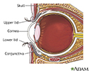
Eye anatomy - illustration
The cornea is the clear layer covering the front of the eye. The cornea works with the lens of the eye to focus images on the retina.
Eye anatomy
illustration
Review Date: 8/20/2016
Reviewed By: Franklin W. Lusby, MD, ophthalmologist, Lusby Vision Institute, La Jolla, CA. Also reviewed by David Zieve, MD, MHA, Isla Ogilvie, PhD, and the A.D.A.M. Editorial team.